Long Bone Drawing
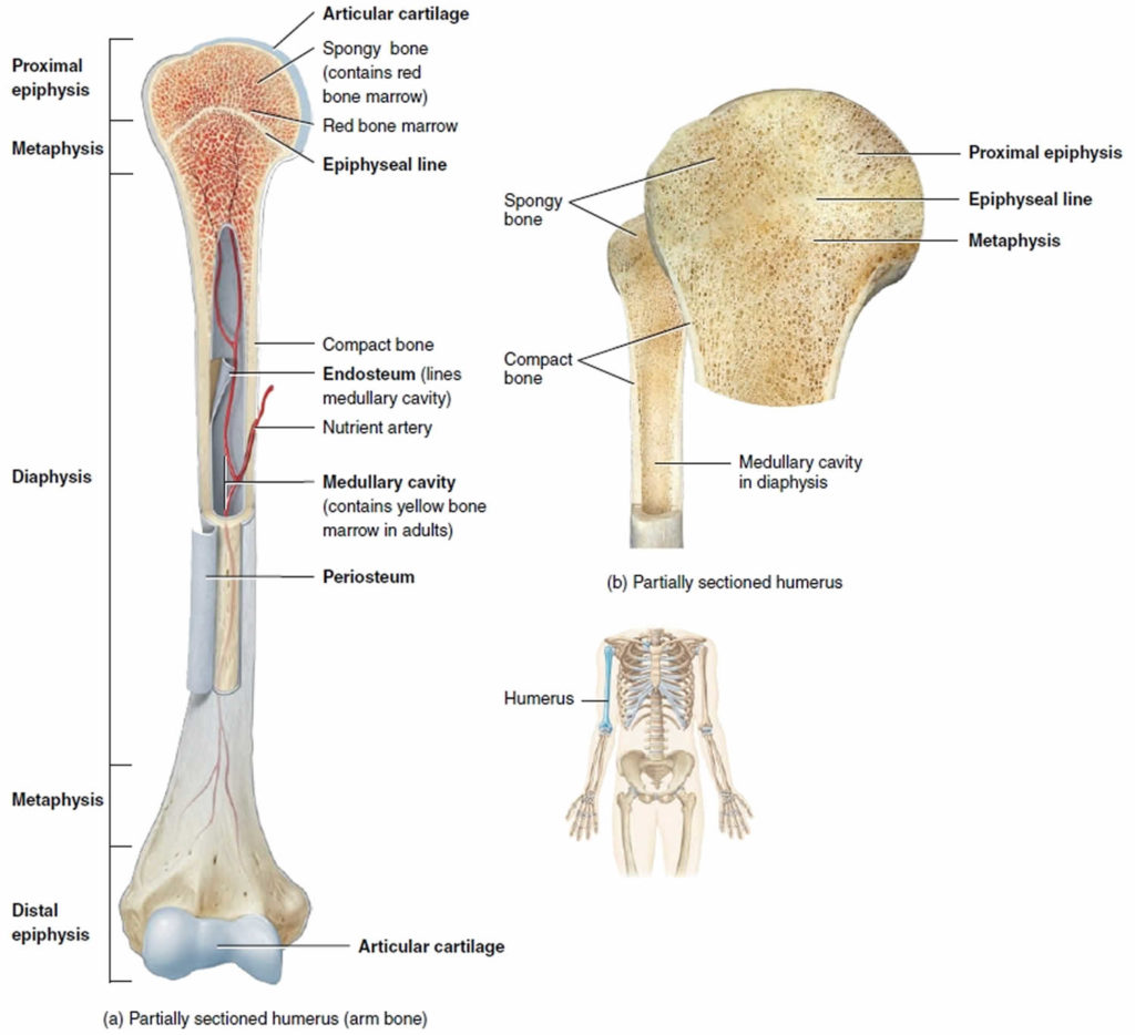
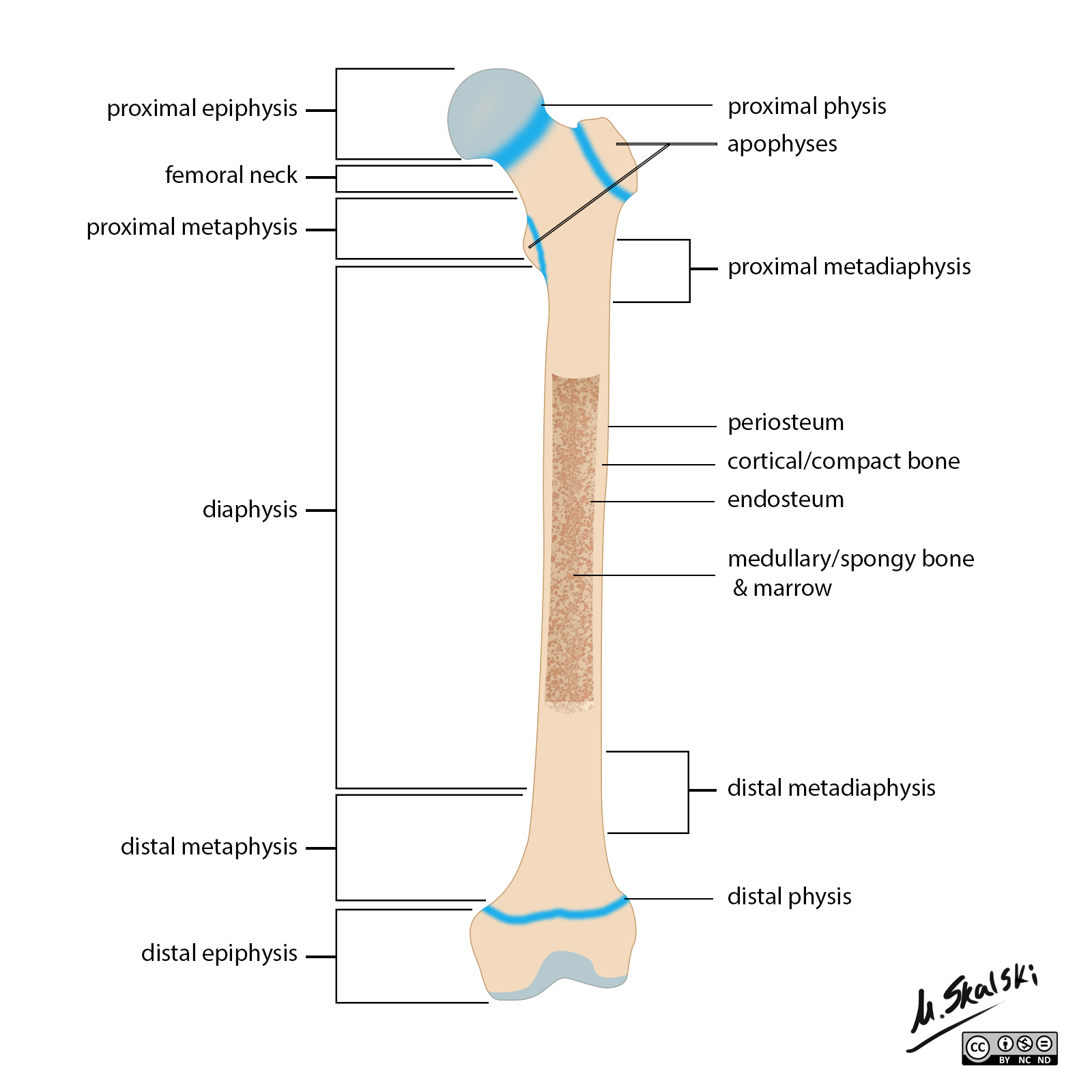
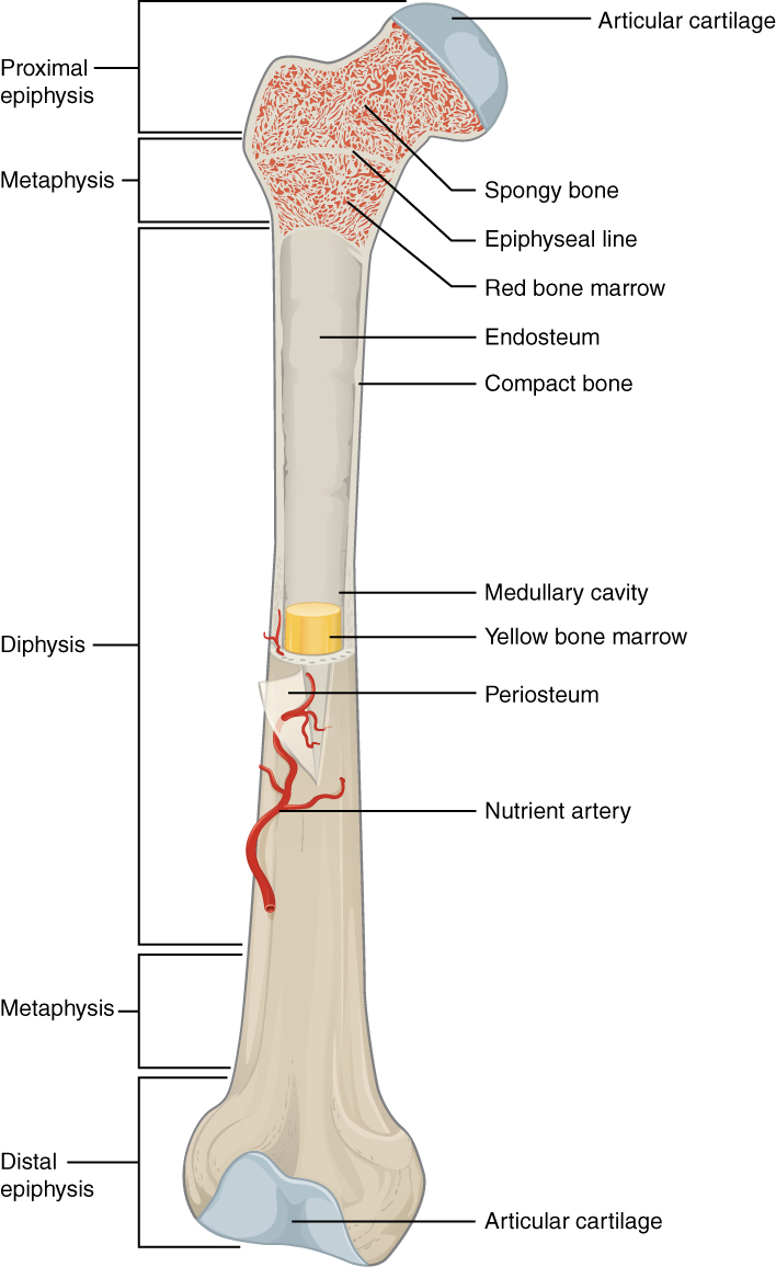
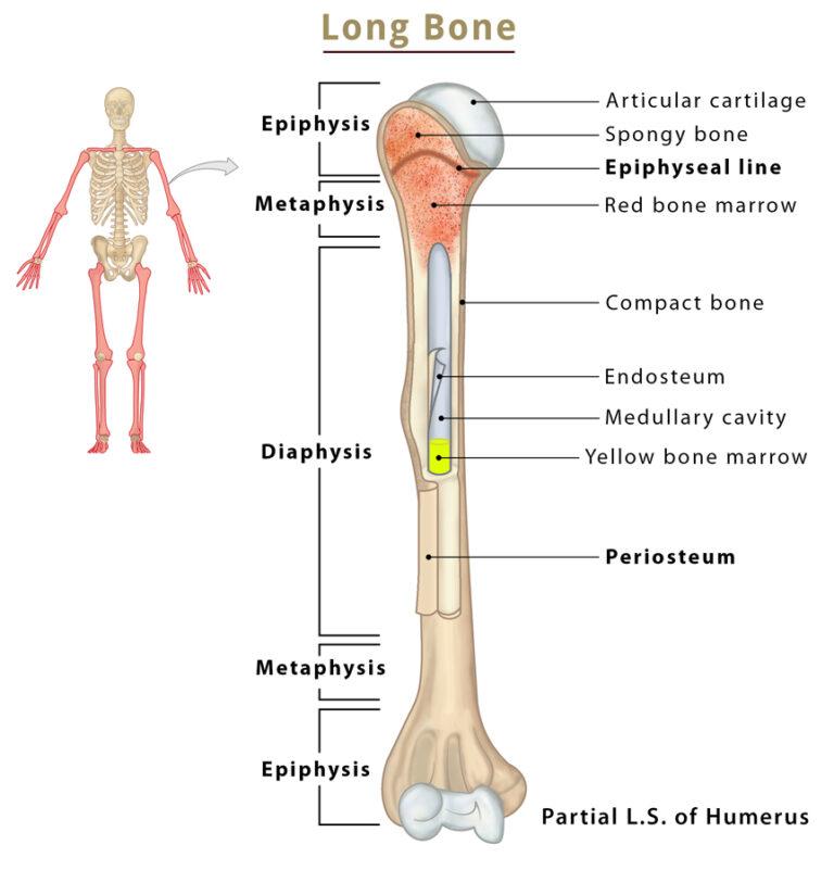
Long Bone Drawing - Web anatomy of a long bone. Then, this line will end in a rounded line at the end. The femur) consists of epiphyses, metaphyses and a diaphysis (shaft). The diaphysis is the hollow, tubular shaft that runs between the proximal and distal ends of the bone. The hollow region in the diaphysis is called the medullary cavity, which is filled with yellow marrow. The clavicle, humerus, radius, ulna, femur, tibia, fibula,. A long bone has two parts: Schematic drawing of a longitudinal section through a long bone (tibia). Web skeletons are strange structures that carry various connotations associated with both life and death. Click the card to flip 👆. The skeletal system 2h 35m. Click the card to flip 👆. Web c = articular cartilage. Anatomy students in traditional classes practice labeling the bone on paper or even doing a coloring activity to help them learn the parts of the bone. The shaft or body or diaphysis of the long bone, and another two are, the extremities. The clavicle, humerus, radius, ulna, femur, tibia, fibula,. Schematic drawing of a longitudinal section through a long bone (tibia). Web anatomy of a long bone. Name the structure of the long bone, at the arrow. The diaphysis and the epiphysis. Web definition of long bones in the body, with a list of names. Their structure & parts (shaft/diaphysis, epiphyses, metaphysis) with labeled diagram The bones belonging to this class are: Web anatomy of a long bone. Web study with quizlet and memorize flashcards containing terms like periosteum, endosteum, articular cartilage and more. The femur) consists of epiphyses, metaphyses and a diaphysis (shaft). The epiphysial plate has been closed in this bone and has become the epiphyseal line after puberty. The diaphysis is the hollow, tubular shaft that runs between the proximal and distal ends of the bone. The diaphysis and the epiphysis. The walls of the diaphysis are composed of dense and. A skeleton drawing can be a great addition to other surreal and fantastical artworks. Click the card to flip 👆. To do this, simply extend another straight line from the end that you drew earlier. G = medullary cavity (yellow marrow) h = endosteum. A long bone has two parts: Long, short, flat, irregular and sesamoid. The hollow region in the diaphysis is called the medullary cavity, which is filled with yellow marrow. The bones belonging to this class are: The diaphysis and the epiphysis ( figure 6.3.1). Web skeletons are strange structures that carry various connotations associated with both life and death. Web a long bone has two main regions: Web study with quizlet and memorize flashcards containing terms like periosteum, endosteum, articular cartilage and more. Web anatomy of a long bone. For this section of your bone drawing, you will be extending the center of the bone on the other side from the one that you drew previously. Web anatomy of. 232k views 5 years ago introduction to anatomy and physiology. Covers the surfaces of bones where they come together to form joints. Web long bone anatomy drawing. The diaphysis and the epiphysis. Click the card to flip 👆. A long bone has two parts: The diaphysis is the tubular shaft that runs between the proximal and distal ends of the bone. The diaphysis and the epiphysis. Study with quizlet and memorize flashcards containing terms like diaphysis, epiphysis. 379k views 9 years ago. The diaphysis is the hollow, tubular shaft that runs between the proximal and distal ends of the bone. They are one of five types of bones: Click the card to flip 👆. The shaft or body or diaphysis of the long bone, and another two are, the extremities. The epiphysial plate has been closed in this bone and has become. The walls of the diaphysis are composed of dense and hard. Click the card to flip 👆. Web c = articular cartilage. A skeleton drawing can be a great addition to other surreal and fantastical artworks. G = medullary cavity (yellow marrow) h = endosteum. Web download scientific diagram | 1: For this section of your bone drawing, you will be extending the center of the bone on the other side from the one that you drew previously. The epiphysial plate has been closed in this bone and has become the epiphyseal line after puberty. The diaphysis and the epiphysis ( figure 6.3.1). The epiphysial plate has been closed in this bone and has become the epiphyseal line after puberty. The human body is a complex, amazing biological machine. They are one of five types of bones: Web the long bones are not straight, but curved, the curve generally taking place in two planes, thus affording greater strength to the bone. Click the card to flip 👆. Long, short, flat, irregular and sesamoid. Web a long bone has two main regions:
Labeled Long Bone Diagram

Long bone anatomy, structure, parts, function and fracture types

Label The Structures Of A Long Bone

Radiopaedia Drawing Anatomy of long bones (femur) English labels

Bone Definition, Anatomy, & Composition Britannica

Long bone Wikipedia

Major Parts Of A Long Bone Diagram

General features of a LONG BONE Biology 225 with Watson at McNeese

Long Bones Anatomy, Examples, Function, & Labeled Diagram

Long Bone Anatomy Drawn & Defined YouTube
232K Views 5 Years Ago Introduction To Anatomy And Physiology.
The Diaphysis Is The Tubular Shaft That Runs Between The Proximal And Distal Ends Of The Bone.
Web The Structure Of A Long Bone Allows For The Best Visualization Of All Of The Parts Of A Bone.
The Diaphysis Is The Hollow, Tubular Shaft That Runs Between The Proximal And Distal Ends Of The Bone.
Related Post: