Veins Of The Arm For Blood Draw
Veins Of The Arm For Blood Draw - (see also vascular access.) indications for venous blood sampling. Lymphedema or dvt in the extremity (choose another extremity) You know which arm we’re talking about. Inspect the patient’s arm for an appropriate venepuncture site: To prevent the vein from rolling, hold the patient’s lower arm and pull the skin below the vein taut. It is a superficial vein located in the cubital fossa, a triangular depression located in the elbow region. Located on the lateral portion of the arm, the cephalic vein is the second most common draw site choice. Also called a blood draw or venipuncture, it’s an important tool for diagnosing many. Web peripheral veins, typically the antecubital veins, are the usual sites for venous blood sampling. This vein is associated with minimal pain and is the most prominent when anchored. Areas of broken, bruised or erythematous skin should be avoided. The main superficial veins of the upper limb include the cephalic and basilic veins. The first step in drawing blood correctly is to identify the appropriate veins to puncture. It is the best because its larger and rolls or moves less than other veins. It is in the inner arm,. The median cubital vein lies between muscles and is usually the most easy to puncture. Every droplet of blood holds a treasure trove of information, from genetic markers to signs of infections or chronic illnesses. Web anchor the vein, then insert the needle quickly to draw blood. The main superficial veins of the upper limb include the cephalic and basilic. Web the median antecubital vein is the most common for blood draws. In most patients, it is very large and easy to access. The median cubital vein in the antecubital fossa is commonly used for venepuncture. Local skin infection, inflammation, trauma or burns. Web locate a vein of a good size that is visible, straight and clear. Unless watching blood leave your body is fun for you, give your arm some privacy. Ultrasound guidance , when equipment and trained personnel are available, can facilitate blood sampling from deep, nonpalpable veins. Lymphedema or dvt in the extremity (choose another extremity) This vein is associated with minimal pain and is the most prominent when anchored. This video is a. (see also vascular access.) indications for venous blood sampling. Local skin infection, inflammation, trauma or burns. It is a superficial vein located in the cubital fossa, a triangular depression located in the elbow region. Every droplet of blood holds a treasure trove of information, from genetic markers to signs of infections or chronic illnesses. Veins of the upper limb are. (see also vascular access.) indications for venous blood sampling. This video is a teaching tutorial for nurs. In this elbow pit, phlebotomists have easy access to the top three vein sites used in phlebotomy: To prevent the vein from rolling, hold the patient’s lower arm and pull the skin below the vein taut. This vein is a gold standard for. The diagram in section 2.3, shows common positions of the vessels, but many variations are possible. It can be subdivided into the superficial system and the deep system. You know which arm we’re talking about. Veins of the upper limb are divided into superficial and deep veins. It is in the inner arm, anterior of the elbow joint. Web phlebotomy, fundamentally, is the act of puncturing a vein to draw blood. Ask the patient to form a fist so the vein becomes more visible, then swiftly insert the needle at a 15 to 30 degree angle. It is the best because its larger and rolls or moves less than other veins. Web the best vein for drawing blood. Web the venous system of the upper limb drains deoxygenated blood from the arm, forearm and hand. Ultrasound guidance , when equipment and trained personnel are available, can facilitate blood sampling from deep, nonpalpable veins. The first step in drawing blood correctly is to identify the appropriate veins to puncture. In this elbow pit, phlebotomists have easy access to the. Use alternate sites (back of hand, forearm) the first area for venipuncture is in the antecubital fossa. Don’t look at that arm. Web the venous system of the upper limb drains deoxygenated blood from the arm, forearm and hand. While it might sound straightforward, the implications and value of this process ripple out far beyond the prick of a needle.. For adult patients, the most common and first choice is the median cubital vein in the antecubital fossa. Inspect the patient’s arm for an appropriate venepuncture site: Any need for a blood sample, usually for various diagnostic tests. Web learn how to find a vein using a tourniquet when drawing blood or starting an iv in the arm (antecubital ac area). Web phlebotomy is when someone uses a needle to take blood from a vein, usually in your arm. Web a blown vein is a vein that’s mildly injured during a blood draw or iv placement. Ultrasound guidance , when equipment and trained personnel are available, can facilitate blood sampling from deep, nonpalpable veins. Web place hot, moist towels over your arms for 10 minutes or so prior to a stick in order to plump up the veins. It is a superficial vein located in the cubital fossa, a triangular depression located in the elbow region. Don’t look at that arm. It can be subdivided into the superficial system and the deep system. Web phlebotomy, fundamentally, is the act of puncturing a vein to draw blood. Web locate a vein of a good size that is visible, straight and clear. The venous system of the upper limb functions to drain deoxygenated blood from the hand, forearm and arm back towards the heart. Veins of the upper limb are divided into superficial and deep veins. Use alternate sites (back of hand, forearm) the first area for venipuncture is in the antecubital fossa.
Diagram Of Veins In Arm For Phlebotomy
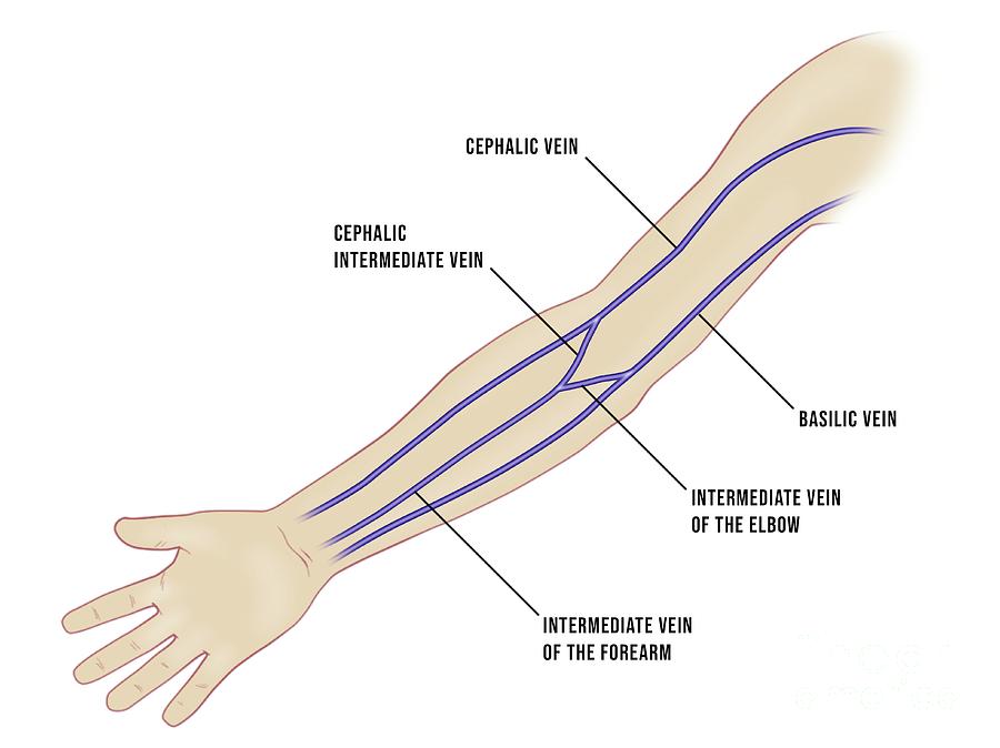
Venous Cannulation Sites In The Arm By Maurizio De Angelis/science

Diagram Of Veins In Arm For Phlebotomy
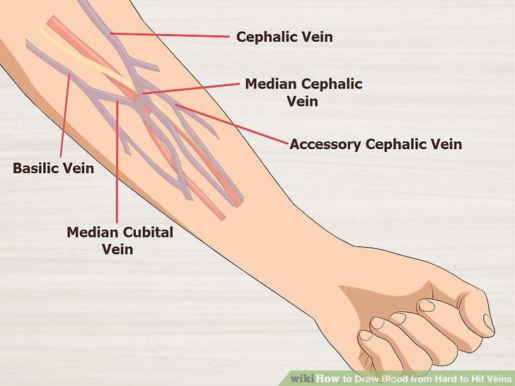
Diagram Of Veins In Arm For Phlebotomy Wiring Diagram Pictures

Diagram Of Veins In Arm For Phlebotomy
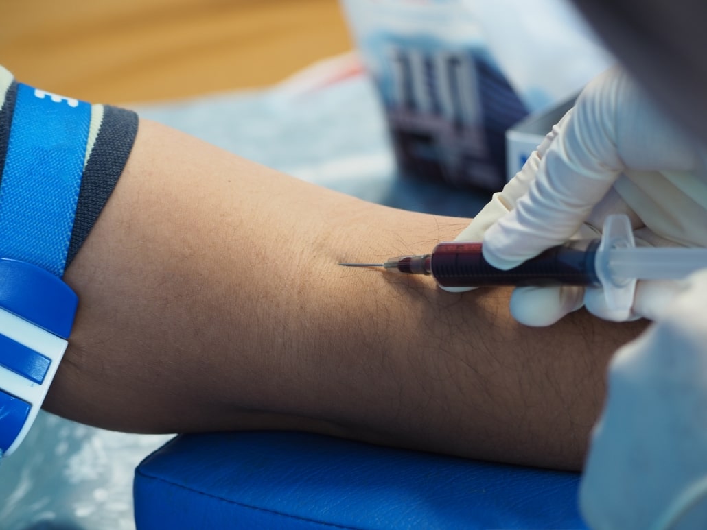
How to draw blood from a patient’s vein as painlessly as possible
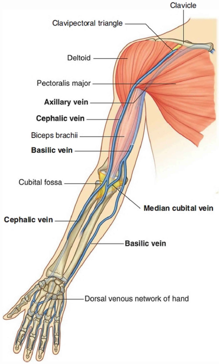
Venipuncture procedure, venipuncture sites, veins & venipuncture
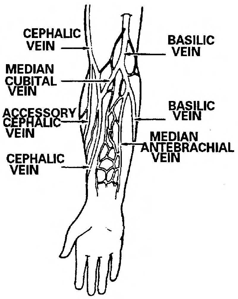
2.3 Procedure for Obtaining a Blood Specimen Intravenous Infusions
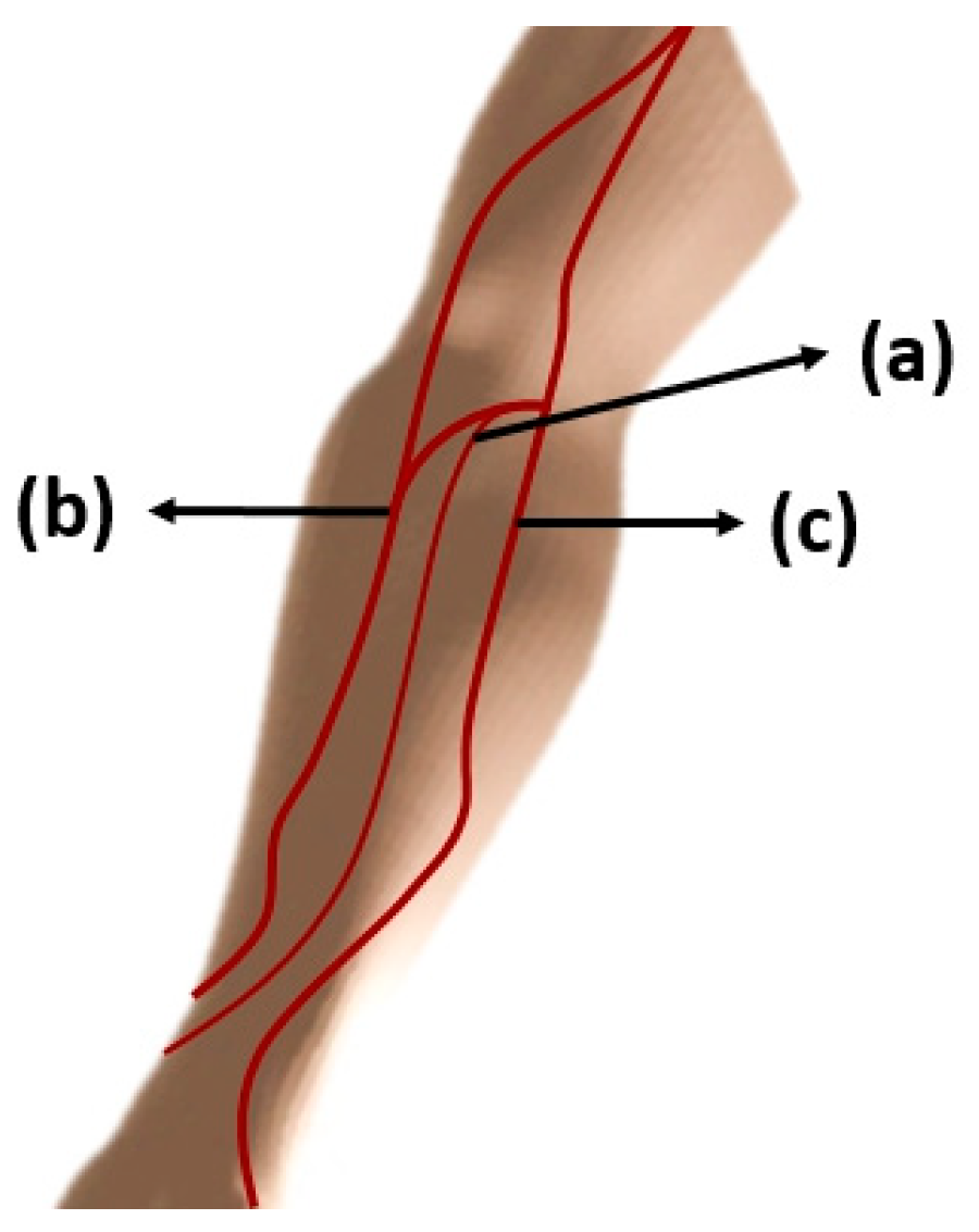
Diagram Of Veins In Arm For Phlebotomy General Wiring Diagram

ns.2018.e10531_0001.jpg
While It Might Sound Straightforward, The Implications And Value Of This Process Ripple Out Far Beyond The Prick Of A Needle.
It Is In The Inner Arm, Anterior Of The Elbow Joint.
It Is The Best Because Its Larger And Rolls Or Moves Less Than Other Veins.
(See Also Vascular Access.) Indications For Venous Blood Sampling.
Related Post: