Smooth Muscle Tissue Drawing
Smooth Muscle Tissue Drawing - Explain how smooth muscles differ from skeletal and cardiac muscles. These muscles are found in almost all organs in the form of bundles or sheaths. Web correlate the microscopic organization of a muscle fiber with the mechanism of contraction and relaxation. Web here presented 43+ smooth muscle drawing images for free to download, print or share. 1 of 3 types of muscle tissue alongside cardiac and skeletal muscle. 80k views 2 years ago class 9 diagram. Web smooth muscle tissue, highlighting the inner circular layer (nuclei then rest of cells in pink), outer longitudinal layer (nuclei then rest of cells), then the serous membrane facing the lumen of the peritoneal cavity. Unlike cardiac and skeletal muscle cells, smooth muscle cells do not exhibit striations since their actin and myosin (thin and thick) protein filaments are not organized as sarcomeres. Web how to draw smooth muscle/muscle tissue diagram/how to draw smooth muscle easy.it is very easy drawing detailed method to help you.i draw the smooth muscle. Web in this article, we'll go through the structure, function, location, characteristics, diagrams and examples of smooth muscle tissue. Web smooth muscle is an essential component of the walls of numerous hollow or tubular organs throughout the body, including blood vessels, airways, and the bladder. Web structure and function. Compare motor and sensory innervation of skeletal muscle tissue. 1 of 3 types of muscle tissue alongside cardiac and skeletal muscle. Smooth muscle is a type of muscle tissue which. Web smooth muscle can be confused with cardiac muscle because the cells are often running in different directions, just as they are in cardiac muscle. Web structure and function. You will also find a details description of the smooth muscle fibers compared to cardiac and skeletal muscles. Skeletal muscle, primate, c.s., h&e. Smooth muscle differs from skeletal muscle in function. Wall of organs like the stomach, oesophagus and intestine. Describe the histological organization of cardiac muscle. Unlike skeletal muscle, smooth muscle is capable of maintaining tone for extended periods and often contracts involuntarily. Web how to draw smooth muscle/muscle tissue diagram/how to draw smooth muscle easy.it is very easy drawing detailed method to help you.i draw the smooth muscle. Learn. Proper physiological functioning of these organs relies heavily on the appropriate activation and contraction of the smooth muscle tissue. Web smooth muscle can be confused with cardiac muscle because the cells are often running in different directions, just as they are in cardiac muscle. Skeletal muscle, primate, c.s., h&e. Smooth muscle cells are a lot smaller than cardiac muscle cells,. Learn how to draw smooth muscle pictures using these outlines or print just for coloring. Proper physiological functioning of these organs relies heavily on the appropriate activation and contraction of the smooth muscle tissue. You will also find a details description of the smooth muscle fibers compared to cardiac and skeletal muscles. Web in this simple guide, i will show. 1 of 3 types of muscle tissue alongside cardiac and skeletal muscle. Explain how smooth muscles differ from skeletal and cardiac muscles. The type of muscle tissue found in the walls of blood vessels and hollow internal organs, such as the stomach, intestine etc. Watch the video tutorial now. The goal of this lab is to learn how to identify. Smooth muscle is widely distributed throughout the body and serves diverse functions. Explain how smooth muscle works with internal organs and passageways through the body. Unlike skeletal muscle, smooth muscle is capable of maintaining tone for extended periods and often contracts involuntarily. The area inside the box is enlarged in the next image. Proper physiological functioning of these organs relies. Web smooth muscle is one of three types of muscle tissue, alongside cardiac and skeletal muscle. Unlike skeletal muscle, smooth muscle is capable of maintaining tone for extended periods and often contracts involuntarily. In relaxed smooth muscle, the nuclei are elongated with rounded ends. You can edit any of drawings via our online image editor before downloading. Smooth muscle is. Wall of organs like the stomach, oesophagus and intestine. The goal of this lab is to learn how to identify and describe the organization and key structural features of smooth and skeletal muscle in sections. By the end of this section, you will be able to: Web smooth muscle tissue, highlighting the inner circular layer (nuclei then rest of cells. Compare motor and sensory innervation of skeletal muscle tissue. Web correlate the microscopic organization of a muscle fiber with the mechanism of contraction and relaxation. Explain how smooth muscles differ from skeletal and cardiac muscles. Watch the video tutorial now. You will also find a details description of the smooth muscle fibers compared to cardiac and skeletal muscles. Web structure and function. Web smooth muscle is an essential component of the walls of numerous hollow or tubular organs throughout the body, including blood vessels, airways, and the bladder. You can edit any of drawings via our online image editor before downloading. Web as you go through these slides, refer to this schematic drawing showing the key structural features and relative sizes of skeletal, smooth, and cardiac muscle as you would observe them with the 40x objective setting. Web smooth muscle is one of three types of muscle tissue, alongside cardiac and skeletal muscle. Explain how smooth muscle differs from skeletal muscle. Smooth muscle is widely distributed throughout the body and serves diverse functions. Explain how smooth muscles differ from skeletal and cardiac muscles. These muscles are found in almost all organs in the form of bundles or sheaths. Nonstriated muscle that serves diverse functions throughout the body and is responsible for involuntary movements. Smooth muscle is a type of muscle tissue which is used by various systems to apply pressure to vessels and organs. Junquiera's basic histology, ch 10: Wall of organs like the stomach, oesophagus and intestine. Smooth muscle differs from skeletal muscle in function. Describe the histological organization of cardiac muscle. How to draw a muscle.
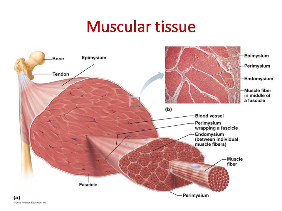
Muscle Tissue Diagram Labeled

Smooth Muscle Tissue Telegraph

LM of a section through human smooth muscle tissue Stock Image P154

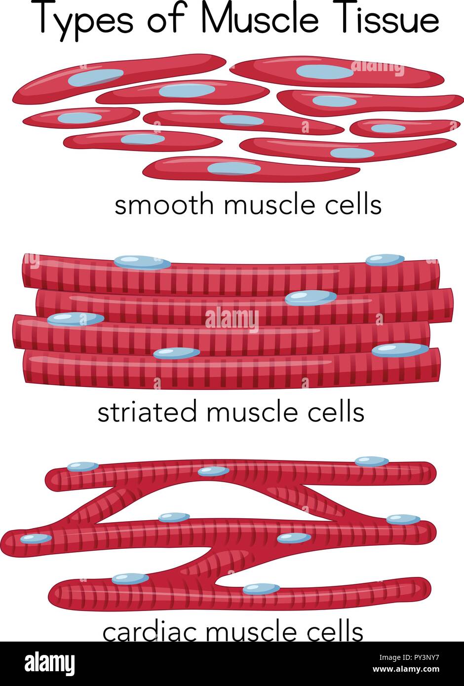
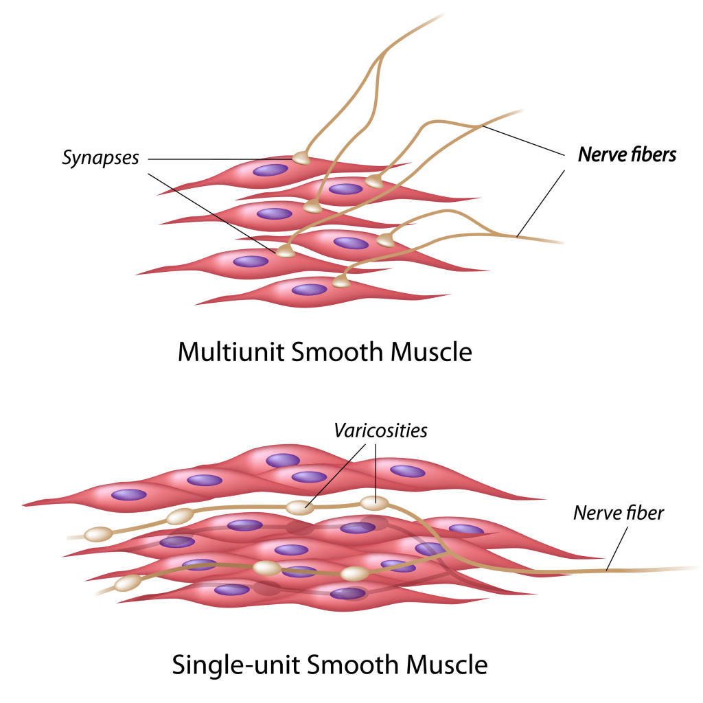
Premium Vector Anatomy of smooth muscle tissue

Smooth Muscle Tissue Diagram Labeled

The Muscular System How We Move Around Interactive Biology, with

Smooth Muscle Diagram Labeled Class 9 Musclenerve Mus vrogue.co

10.8 Smooth Muscle Douglas College Human Anatomy and Physiology I

Smooth Muscle Tissue Diagram Drawing ezildaricci

How To Draw Muscle Cell Step by Step YouTube
Web Here Presented 43+ Smooth Muscle Drawing Images For Free To Download, Print Or Share.
Web Smooth Muscle Can Be Confused With Cardiac Muscle Because The Cells Are Often Running In Different Directions, Just As They Are In Cardiac Muscle.
To Find The Muscle Layer, Look At The At Slide At The Lowest Power (This Is About The Same As Looking At The Glass Slide With The Naked Eye).
Web Correlate The Microscopic Organization Of A Muscle Fiber With The Mechanism Of Contraction And Relaxation.
Related Post: