Simple Cuboidal Epithelium Drawing
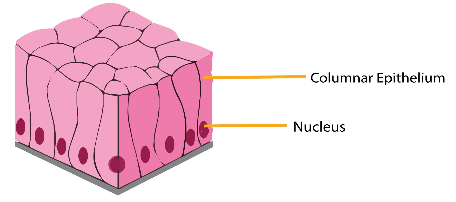
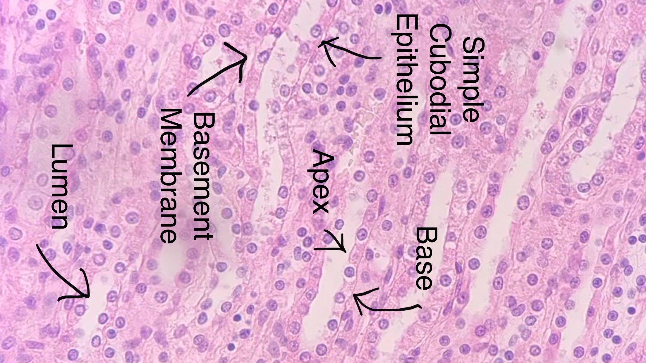
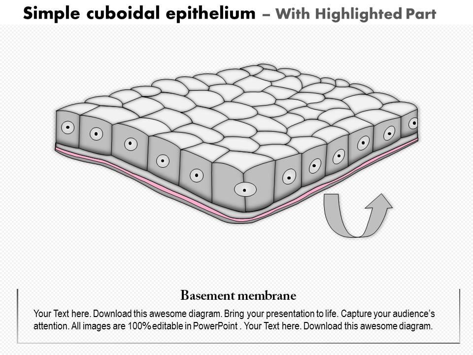
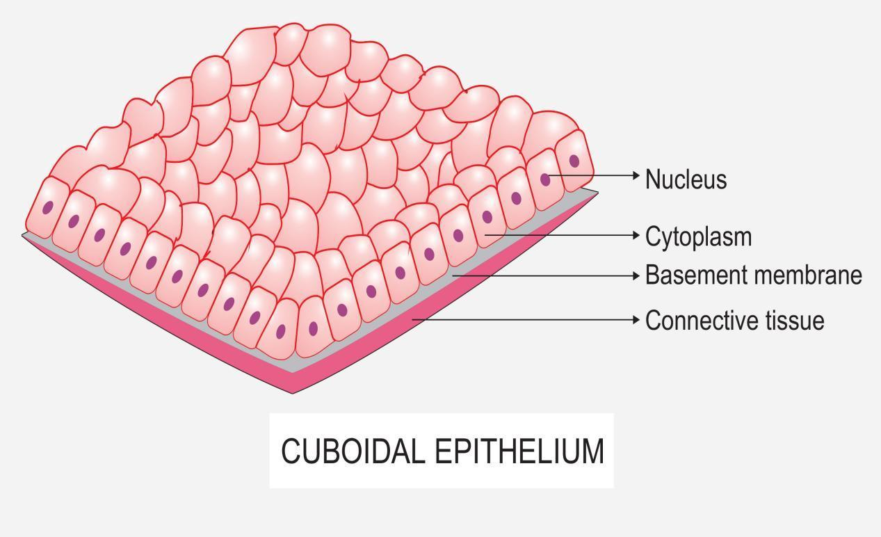
Simple Cuboidal Epithelium Drawing - 713 views 2 years ago. Structure of the simple cuboidal epithelium. Simple cuboidal epithelia are observed in the lining of the kidney tubules and in the ducts of glands. Explain the structure and function of epithelial tissue. These cuboidal cells have large, spherical and central nuclei. With large, rounded, centrally located nuclei, all the cells of this epithelium are directly attached to the basement membrane. Secrets lubricating substance, allows diffusion and filtration. While both figures show simple cuboidal epithelium of the renal tubules in the kidney, figure 4 shows a cross section and figure 5 shows a longitudinal section. Types of simple cuboidal epithelia Web table of contents. Web human structure virtual microscopy. Overview and types of epithelial tissue. Web simple cuboidal epithelia occur widely in the body in many glands and glandular ducts, such as the salivary ducts, pancreatic duct, bile duct, and kidney tubules.these epithelia are composed of cells that are short prisms with a top, bottom, and six sides. Simple epithelium [13:17] structures and types. This is the 2nd video of our series.in which we are making you learn how to draw simple cuboidal epithelium.if you like our video.give a thumbs. Methods and types of secretion. Web function and classes. Web drawing histological diagram of simple cuboidal epithelia.useful for all medical students.drawn by using h & e pencils.explanation on epithelia while drawing. Web there are. The proximal convoluted tubules of the kidney). These epithelia are involved in the secretion and absorptions of molecules requiring active transport. Structure of the simple cuboidal epithelium. Web welcome to diya's art tutorial youtube channel today in this video i'm showing how to draw cuboidal. A columnar epithelial cell looks like a column or a tall rectangle. Simple cuboidal epithelia are observed in the lining of the kidney tubules and in the ducts of glands. Web human structure virtual microscopy. Web welcome to diya's art tutorial youtube channel today in this video i'm showing how to draw cuboidal. The proximal convoluted tubules of the kidney). Simple cuboidal epithelia are observed in the lining of the kidney tubules. This is the 2nd video of our series.in which we are making you learn how to draw simple cuboidal epithelium.if you like our video.give a thumbs. Overview and types of epithelial tissue. 713 views 2 years ago. Simple cuboidal epithelia are observed in the lining of the kidney tubules and in the ducts of glands. These epithelia are involved in. Web there are three basic shapes used to classify epithelial cells. Web human structure virtual microscopy. Web simple cuboidal epithelium to help you understand how to identify simple squamous epithelium, we have included two examples of this tissue. Web simple cuboidal epithelia occur widely in the body in many glands and glandular ducts, such as the salivary ducts, pancreatic duct,. Web function and classes. 713 views 2 years ago. Secrets lubricating substance, allows diffusion and filtration. Structure of the simple cuboidal epithelium. Web simple cuboidal epithelia occur widely in the body in many glands and glandular ducts, such as the salivary ducts, pancreatic duct, bile duct, and kidney tubules.these epithelia are composed of cells that are short prisms with a. Web simple cuboidal epithelium to help you understand how to identify simple squamous epithelium, we have included two examples of this tissue. Jana vasković, md • reviewer: Types of simple cuboidal epithelia When viewed from atop these cells are square in shape. Secrets lubricating substance, allows diffusion and filtration. These cuboidal cells have large, spherical and central nuclei. Blood and lymphatic vessels, air sacs of lungs, lining of the heart. These epithelia are active in the secretion and absorptions of molecules. Location and examples of simple cuboidal epithelium. Web table of contents. The proximal convoluted tubules of the kidney). When viewed from atop these cells are square in shape. Use the image slider below to learn how to use a microscope to identify and study simple cuboidal epithelium on a. These epithelia are active in the secretion and absorption of molecules. Jana vasković, md • reviewer: Web function and classes. A cuboidal epithelial cell looks close to a square. Web welcome to diya's art tutorial youtube channel today in this video i'm showing how to draw cuboidal. A squamous epithelial cell looks flat under a microscope. Explain the structure and function of epithelial tissue. Use the image slider below to learn more about the characteristics of simple cuboidal epithelium. Web simple cuboidal epithelia occur widely in the body in many glands and glandular ducts, such as the salivary ducts, pancreatic duct, bile duct, and kidney tubules.these epithelia are composed of cells that are short prisms with a top, bottom, and six sides. Secrets lubricating substance, allows diffusion and filtration. These epithelia are involved in the secretion and absorptions of molecules requiring active transport. These epithelia are active in the secretion and absorptions of molecules. The proximal convoluted tubules of the kidney). Web simple cuboidal epithelium to help you understand how to identify simple squamous epithelium, we have included two examples of this tissue. 713 views 2 years ago. With large, rounded, centrally located nuclei, all the cells of this epithelium are directly attached to the basement membrane. Simple cuboidal epithelia are observed in the lining of the kidney tubules and in the ducts of glands. A columnar epithelial cell looks like a column or a tall rectangle.
Simple Cuboidal Epithelial Tissue Diagram

Simple cuboidal epithelium Diagram Quizlet

Simple Cuboidal Epithelium Labeled Cell Membrane Drawo

Simple cuboidal epithelium

0614 Simple Cuboidal Epithelium Medical Images For Powerpoint

Simple cuboidal epithelium

0614 Simple Cuboidal Epithelium Medical Images For Powerpoint

How To Draw Cuboidal Epithelial Tissue (step by step) how_to_draw

How to draw stratified cuboidal epithelium easy way YouTube

Simple Cuboidal Epithelium Diagram
Overview And Types Of Epithelial Tissue.
These Epithelia Are Active In The Secretion And Absorption Of Molecules.
Web There Are Three Basic Shapes Used To Classify Epithelial Cells.
This Is The 2Nd Video Of Our Series.in Which We Are Making You Learn How To Draw Simple Cuboidal Epithelium.if You Like Our Video.give A Thumbs.
Related Post: