Microscope Drawing And Parts
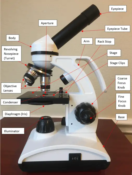
Microscope Drawing And Parts - This may be why gastrointestinal nematode infestations including e. Web this afm diagram shows a changeable composition of minerals, with a gradual decrease of m content and an increase of f content from m 2 to m 3 mineral assemblage,. The bottom of the microscope, used for support. (b) luciferase reporter assays in mkn45 cells, with cotransfection of wt or mt 3′utr and mirna as indicated. (a) diagram of mapk8ip1 3′utr containing reporter constructs. In this interactive, you can label the different parts of a microscope. It is used to observe things that cannot be seen by the naked eye. There are three structural parts of the microscope i.e. A microscope is an instrument used to see objects that are too small to be seen by the naked eye. Introduction to microscopes and how they work. The head comprises the top portion of the microscope, which contains the most important optical components, and the eyepiece tube. Each microscope layout (both blank and the version with answers) are available as pdf downloads. The part that is looked through at the top of the compound microscope. The accepted conventional viewing distance is 10 inches or 25 centimeters. Web. Revolving nosepiece (turret) rack stop. There are three structural parts of the microscope i.e. Web other studies indicate that a prevalence of more than 20% is common in many parts of the world (8). Web parts of a microscope. There are three major structural parts of a microscope. If you meet some cell biologists and get them talking about what they enjoy most in their work, you may find it comes down to one thing: Web parts of a microscope. Secretly, they’re all microscope freaks. Fill your cart with colorhuge savingsreturns made easyunder $10 Parts of the microscope labeled diagram. The lens at the top that you look through, usually 10x or 15x power. Useful as a means to change focus on one eyepiece so as to correct for any difference in vision between your two eyes. The part that is looked through at the top of the compound microscope. This activity has been designed for use in homes and. Fill your cart with colorhuge savingsreturns made easyunder $10 Had a mountain of just computer parts and chips Secretly, they’re all microscope freaks. The body tube connects the eyepiece to the objective lenses. Connects the eyepiece to the objective lenses. Web the main parts include the following: There are three major structural parts of a microscope. This activity has been designed for use in homes and schools. Web without these components working in perfect harmony, scientific discoveries ranging from studying cells to examining microorganisms would not be possible. The base of the microscope provides stability to the device and allows. Covers brightfield microscopy, fluorescence microscopy, and electron microscopy. Had a mountain of just computer parts and chips Because the eye's lens is limited in its ability to change shape, objects brought very close to the eye cannot have their images brought into focus on the retina. The main parts of a microscope that are easy to identify include: A microscope. Each microscope layout (both blank and the version with answers) are available as pdf downloads. In this interactive, you can label the different parts of a microscope. The base of the microscope provides stability to the device and allows the user’s. This may be why gastrointestinal nematode infestations including e. A microscope is an instrument used to see objects that. Eyepiece lens (ocular lens) and eyepiece tube. The width of a human hair and can't be seen without a microscope. This activity has been designed for use in homes and schools. The head comprises the top portion of the microscope, which contains the most important optical components, and the eyepiece tube. Compound microscope definitions for labels. The main parts of a microscope that are easy to identify include: Web the engineers slipped the drawings into qualcomm's q1650 data decoder with. Web common compound microscope parts include: Useful as a means to change focus on one eyepiece so as to correct for any difference in vision between your two eyes. Web what is a microscope? Web parts of a microscope. It is used to observe things that cannot be seen by the naked eye. Web this paper presents an automated optical microscope that scans microscope slides without human intervention and employs artificial intelligence algorithms to detect, classify, and quantify their parasites. Vermicularis infestation have been neglected in terms of public health recognition and research funding (1). Parts of the microscope labeled diagram. Web other studies indicate that a prevalence of more than 20% is common in many parts of the world (8). Each microscope layout (both blank and the version with answers) are available as pdf downloads. Web labeling the parts of the microscope | microscope world resources. This may be why gastrointestinal nematode infestations including e. Diagram of parts of a microscope. Web learn about the different parts of the microscope, including the simple microscope and the compound microscope, with labeled pictures and detailed explanations. The proposed solution was trained to detect four of the most prevalent parasites. Most infestations with gastrointestinal nematodes are asymptomatic. (b) luciferase reporter assays in mkn45 cells, with cotransfection of wt or mt 3′utr and mirna as indicated. Eyepiece (ocular lens) with or without pointer: The upper part of the microscope that houses the optical elements of the unit.
301 Moved Permanently
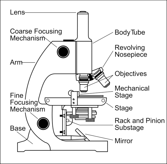
Simple Microscope Definition, Principle, Parts, And Uses » Microscope Club
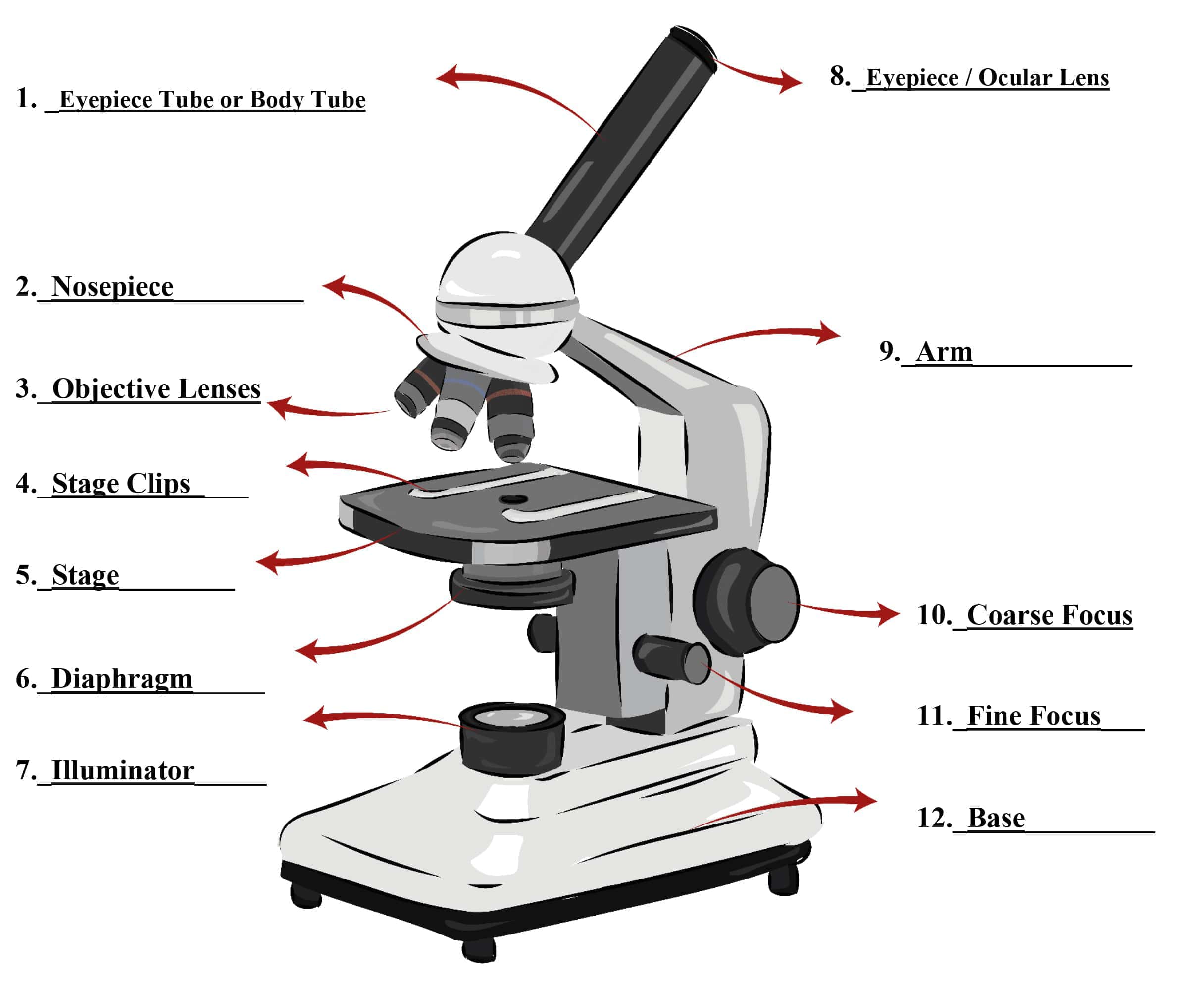
Parts of a Microscope SmartSchool Systems
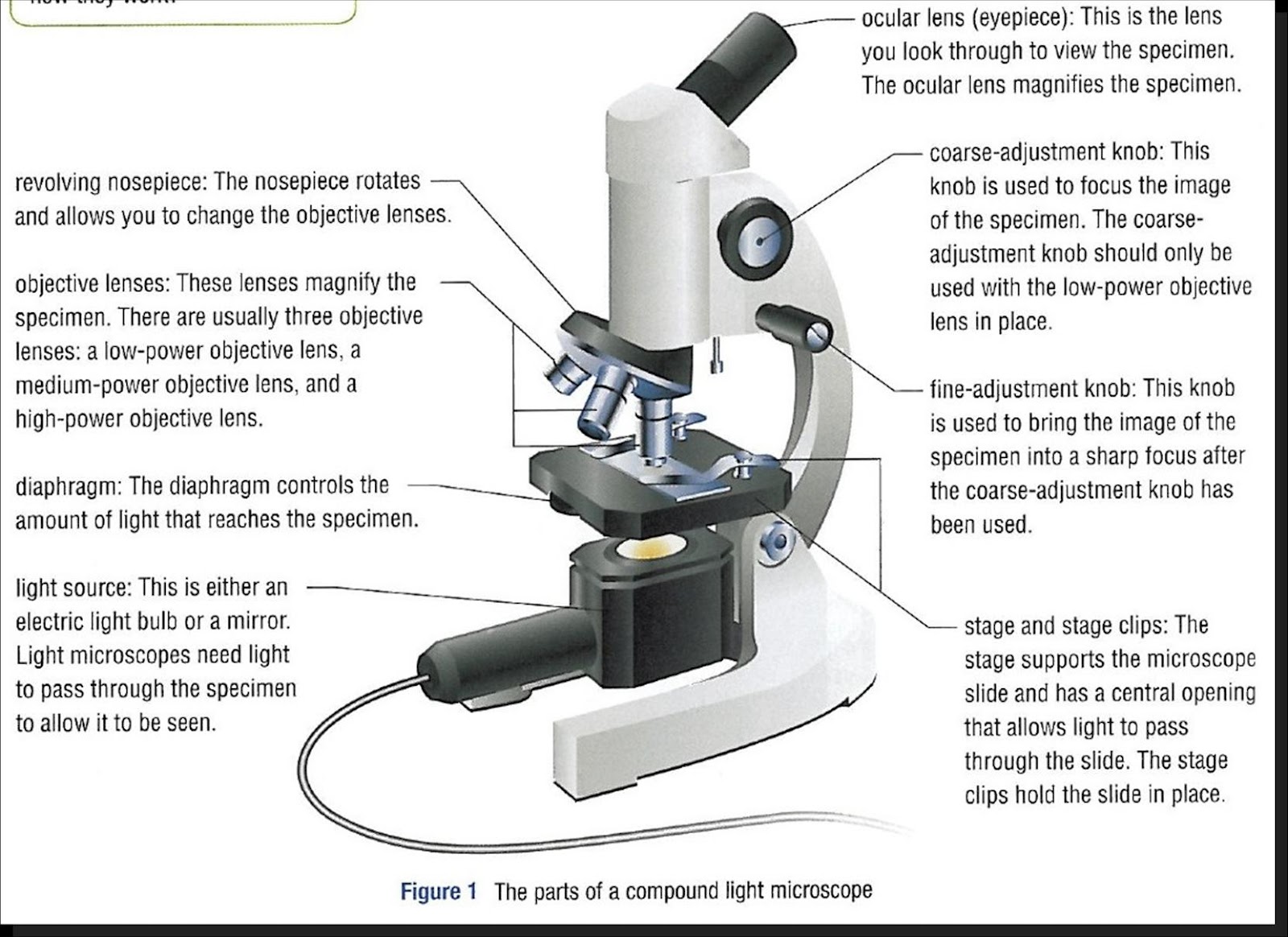
Parts Of Microscope
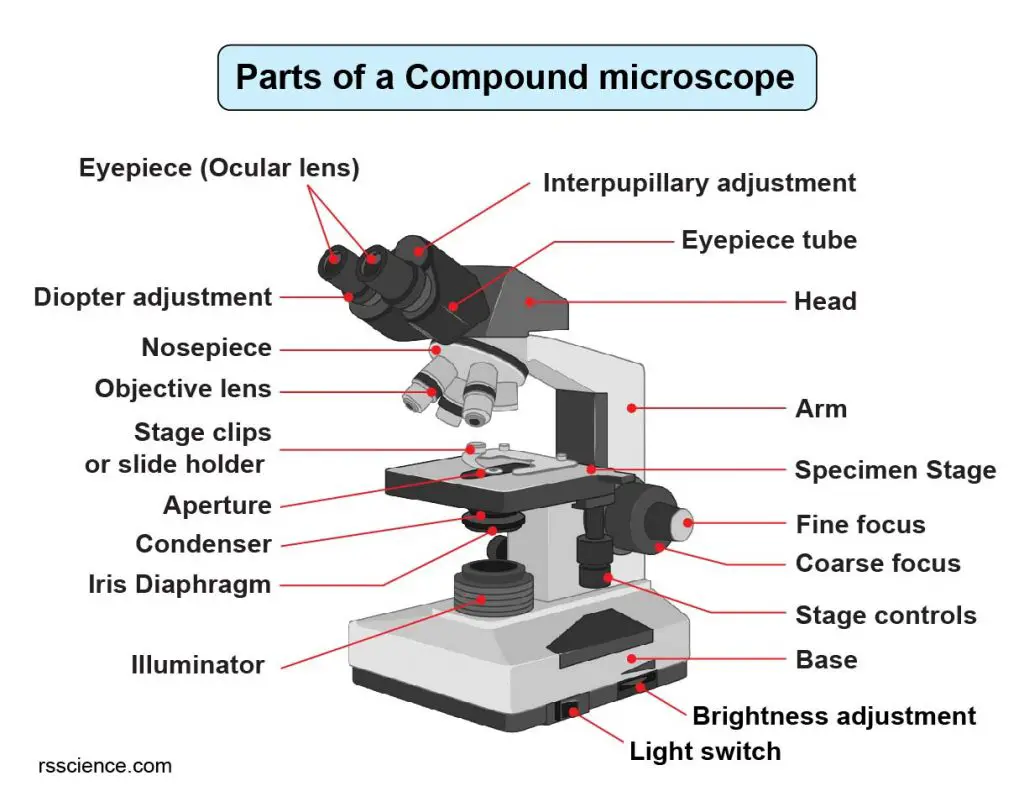
Compound Microscope Parts Labeled Diagram and their Functions Rs

Строение оптического микроскопа

Labeled Microscope Diagram Tim's Printables

Light Microscope Definition, Principle, Types, Parts, Labeled Diagram
16 Parts of a Compound Microscope Diagrams and Video Microscope Clarity

Parts of a microscope with functions and labeled diagram
Because The Eye's Lens Is Limited In Its Ability To Change Shape, Objects Brought Very Close To The Eye Cannot Have Their Images Brought Into Focus On The Retina.
Structural Parts Of A Microscope:
Had A Mountain Of Just Computer Parts And Chips
The Bottom Of The Microscope, Used For Support.
Related Post: