Hyaline Cartilage Drawing
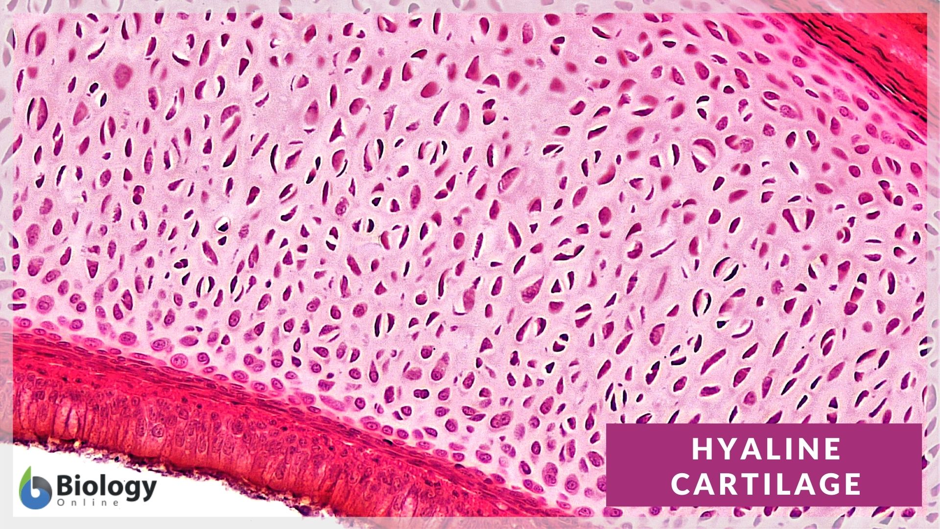
Hyaline Cartilage Drawing - Web the hyaline cartilage in the trachea is in the middle of the tracheal wall. Isogenous groups and interstitial growth results when chondrocytes divide and produce extracellular matrix. Use the image slider below to learn more about the characteristics of hyaline cartilage. Web micrograph showing fibrocartilage (centre) surrounded by areas of hyaline cartilage (upper left and right) that are being converted to bone. These cells have relatively small nuclei and often demonstrate lipid. Hyaline cartilage is the most prevalent type, forming articular cartilages and the framework for parts of the nose, larynx, and trachea. It contains no nerves or blood vessels, and its structure is relatively simple. Cartilage is flexible connective tissue found throughout the whole body. Cells that form and maintain the cartilage. Star star star star star. Territorial matrix lies immediately around each isogenous group and is high in glycosaminoglycans. This image shows a cross section of a cartilage ring that supports the trachea and maintains the. Hyaline cartilage is the most prevalent type, forming articular cartilages and the framework for parts of the nose, larynx, and trachea. Isogenous groups and interstitial growth results when chondrocytes divide. Hyaline cartilage is the most prevalent type, forming articular cartilages and the framework for parts of the nose, larynx, and trachea. Web during embryonic development, hyaline cartilage serves as temporary cartilage models that are essential precursors to the formation of most of the axial and appendicular skeleton. You can begin to see the details in hyaline cartilage (hc. Use the. Web hyaline cartilage is the most common of the three types of cartilage. Web the hyaline cartilage in the trachea is in the middle of the tracheal wall. Hyaline cartilage is a type of connective tissue found in areas such as the nose, ears, and trachea of the human body. A type of cartilage found on many joint surfaces; These. This article will focus on important features of hyaline cartilage, namely its matrix, chondrocytes, and perichondrium. Note the numerous chondrocytes in this image, each located within lacunae and surrounded by the cartilage they have produced. Hyaline cartilage is a type of connective tissue found in areas such as the nose, ears, and trachea of the human body. [digitalscope] note the. Hyaline cartilage is the most prevalent type, forming articular cartilages and the framework for parts of the nose, larynx, and trachea. A joint of the jaw that connects it to the temporal bones of the skull. The bar shows the position of the hyaline cartilage. Territorial matrix lies immediately around each isogenous group and is high in glycosaminoglycans. Web lab. This image shows a cross section of a cartilage ring that supports the trachea and maintains the. A joint of the jaw that connects it to the temporal bones of the skull. Web the illustrative book of cartilage repair. Cells that form and maintain the cartilage. It is also most commonly found in the ribs, nose, larynx, and trachea. Web hyaline cartilage tissue (also referred to as hyaline connective tissue or hyaline tissue) is a type of a cartilage tissue. A joint of the jaw that connects it to the temporal bones of the skull. Web likecomment share subscribe #hyalinecartilage #histodiagrams #hyalinecartilagediagram #cartilagehistology Hyaline cartilage is the most widespread and is the type that makes up the embryonic skeleton.. Note the numerous chondrocytes in this image, each located within lacunae and surrounded by the cartilage they have produced. It contains no nerves or blood vessels, and its structure is relatively simple. Use the image slider below to learn more about the characteristics of hyaline cartilage. Star star star star star. This post will describe the basic histology of hyaline. We will examine those tissues in greater detail in lab 5 the appendicular skeleton & lab 6 the axial skeleton. A type of cartilage found on many joint surfaces; Supporting connective tissue comprises bone and cartilage. Tamás oláh, tunku kamarul, henning madry & malliga raman murali. You can begin to see the details in hyaline cartilage (hc. This post will describe the basic histology of hyaline cartilage with slide images and labeled diagram. A joint of the jaw that connects it to the temporal bones of the skull. Multipotential cells in the fibrous layer of the perichondrium differentiate into chondroblasts in the chondrogenic layer. It contains no nerves or blood vessels, and its structure is relatively simple.. Hyaline cartilage is a type of connective tissue found in areas such as the nose, ears, and trachea of the human body. Hyaline cartilage is the most prevalent type, forming articular cartilages and the framework for parts of the nose, larynx, and trachea. Web hyaline cartilage is the most common of the three types of cartilage. Tamás oláh, tunku kamarul, henning madry & malliga raman murali. Web the illustrative book of cartilage repair. Step by step drawing of histology of hyaline cartilage Where is hyaline cartilage found? It contains no nerves or blood vessels, and its structure is relatively simple. You can begin to see the details in hyaline cartilage (hc. We will examine those tissues in greater detail in lab 5 the appendicular skeleton & lab 6 the axial skeleton. Use the image slider below to learn how to use a microscope to identify and study hyaline cartilage on a microscope slide of the trachea. Use the image slider below to learn more about the characteristics of hyaline cartilage. New articular cartilage is limited to interstitial growth because of the absence of a perichondrium. A type of cartilage found on many joint surfaces; Star star star star star. Web in part i , there are four slides to examine, showing hyaline cartilage (webslide 26), elastic cartilage (webslide 12 and umich 44h), and fibrocartilage (webslides 45 and 74).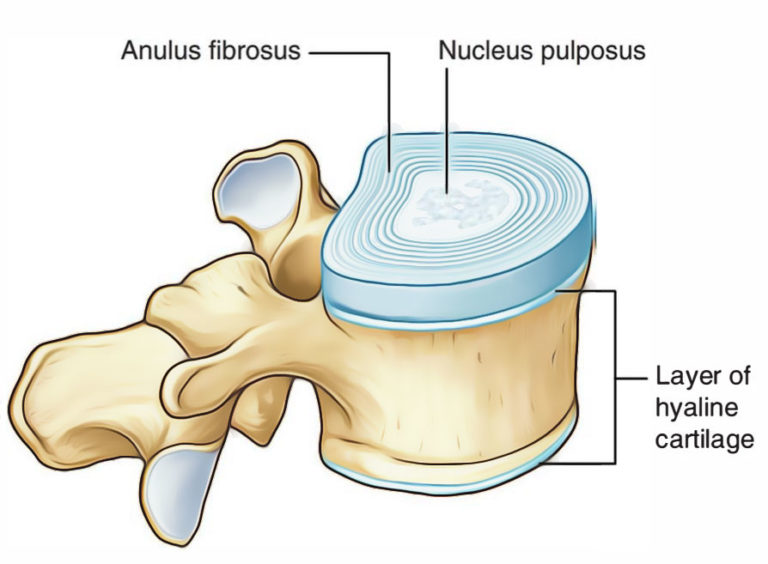
Hyaline Cartilage Earth's Lab

Illustrations Hyaline Cartilage General Histology
Hyaline cartilage hires stock photography and images Alamy

How to Draw Hyaline Cartilage Simple and easy steps Biology Exam
Hyaline Cartilage Cells ClipArt ETC

Hyaline Cartilage Labeled Diagram

Hyaline Cartilage Drawing YouTube
Hyaline cartilage Definition and Examples Biology Online Dictionary

Schematic drawing of articular (hyaline) cartilage containing abundant

Histology Image Cartilage
Articular Cartilage Contains No Blood Vessels Or Nerves.
Web Lab 3 Exercise 3.3.1 3.3.
Web The Hyaline Cartilage In The Trachea Is In The Middle Of The Tracheal Wall.
When A Chondroblast Has Surrounded Itself With Cartilage, It Is Then Called A Chondrocyte.
Related Post: