Human Heart Drawing Labeled
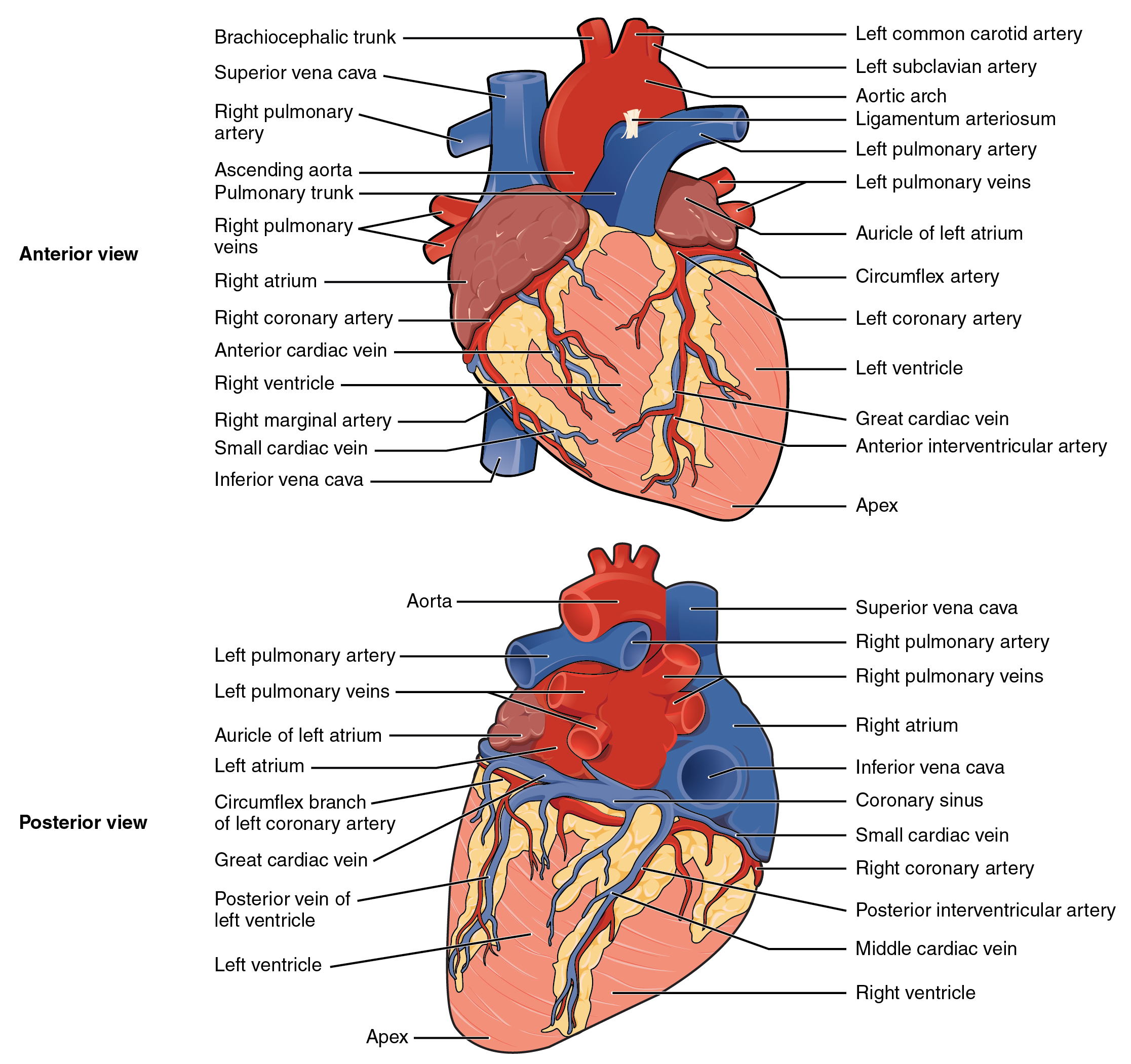
Human Heart Drawing Labeled - Web + show all. The heart is made up of four chambers: Web anatomy of the human heart and coronaries: Web your heart’s main function is to move blood throughout your body. Web inside, the heart is divided into four heart chambers: It rests on the diaphragm, the muscular partition between the chest and the abdominal cavity. In this lecture, dr mike shows the two best ways to draw and label the heart!. The two upper chambers are called the left and the right atria, and the two lower chambers are known as the left and the right ventricles. Click to view large image. Web anatomy of the human heart. The two upper chambers are called the left and the right atria, and the two lower chambers are known as the left and the right ventricles. Shading the lower sections of the heart. Web the heart is made of three layers of tissue. The heart is a mostly hollow, muscular organ composed of cardiac muscles and connective tissue that acts.. They will be to the lower left of the aorta. In humans, the heart is situated between the two lungs and slightly to the left of center, behind the breastbone. The human heart is located within the thoracic cavity, medially between the lungs in the space known as the mediastinum. Web inside, the heart is divided into four heart chambers:. New 3d rotate and zoom. The heart wall is made up of three layers: Find a piece of paper and something to draw with. This tool provides access to several medical illustrations, allowing the user to interactively discover heart anatomy. Web dr matt & dr mike. If you're trying to identify parts of the heart for a class or just for fun, consider adding the names of each segment. The human heart, comprises four chambers: Shading the lower sections of the heart. Click to view large image. New 3d rotate and zoom. Find an image that displays the entire heart, and click on it to enlarge it. Web this will teach you how to draw human heart diagram easily. Light pencil shading of the heart. The two upper chambers are called the atria, the remaining two lower chambers are the ventricles. Controls the rhythm and speed of your heart rate. The heart is a mostly hollow, muscular organ composed of cardiac muscles and connective tissue that acts. The middle layer of the heart wall is called myocardium. The upper two chambers of the heart are called auricles. Once you’re feeling confident, you can test yourself using the unlabeled diagrams of the parts of the heart below. [right atrium and ventricle. Endocardium is the thin inner lining of the heart chambers and also forms the surface of the valves. Web function and anatomy of the heart made easy using labeled diagrams of cardiac structures and blood flow through the atria, ventricles, valves, aorta, pulmonary arteries veins, superior inferior vena cava, and chambers. Right atrium, left atrium, right ventricle and left ventricle.. To find a good diagram, go to google images, and type in the internal structure of the human heart. The lower two chambers of the heart are called ventricles. Find a piece of paper and something to draw with. Learn all about the heart, blood vessels, and composition of blood itself with our 3d models and explanations of cardiovascular system. Light pencil shading of the heart. Web this will teach you how to draw human heart diagram easily. Light sketching of the heart. The two upper chambers are called the atria, the remaining two lower chambers are the ventricles. In this interactive, you can label parts of the human heart. Web the cardiovascular system. The right and left sides of the heart are separated by a muscle called the “septum.”. [right atrium and ventricle of the heart (labeled)] Learn all about the heart, blood vessels, and composition of blood itself with our 3d models and explanations of cardiovascular system anatomy and physiology. Both sides work together to efficiently circulate the. Includes an exercise, review worksheet, quiz, and model drawing of an anterior vi In this interactive, you can label parts of the human heart. 41k views 1 year ago cardiovascular system. Learn all about the heart, blood vessels, and composition of blood itself with our 3d models and explanations of cardiovascular system anatomy and physiology. Web dr matt & dr mike. Web function and anatomy of the heart made easy using labeled diagrams of cardiac structures and blood flow through the atria, ventricles, valves, aorta, pulmonary arteries veins, superior inferior vena cava, and chambers. Shading the upper sections of the heart in pen. Once you’re feeling confident, you can test yourself using the unlabeled diagrams of the parts of the heart below. The human heart, comprises four chambers: Find a piece of paper and something to draw with. To find a good diagram, go to google images, and type in the internal structure of the human heart. The right and left sides of the heart are separated by a muscle called the “septum.”. Start with the pulmonary veins. Right atrium, left atrium, right ventricle and left ventricle. Selecting or hovering over a box will highlight each area in the diagram. The heart is a mostly hollow, muscular organ composed of cardiac muscles and connective tissue that acts.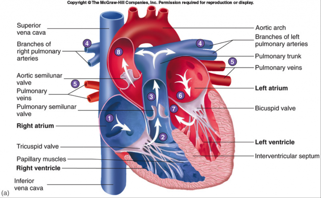
Human HeartGross structure and Anatomy Online Biology Notes
Heart Anatomy Labeled Diagram, Structures, Blood Flow, Function of

The 18 parts of the human heart, and their functions Wellnessbeam

FileHeart diagramen.svg Wikipedia
.svg/1043px-Diagram_of_the_human_heart_(cropped).svg.png)
FileDiagram of the human heart (cropped).svg Wikipedia
19.1 Heart Anatomy Anatomy and Physiology

Anatomy of the human heart

Heart Anatomy chambers, valves and vessels Anatomy & Physiology

How to Draw the Internal Structure of the Heart 14 Steps

Basic Anatomy of the Human Heart Cardiology Associates of Michigan
If You're Trying To Identify Parts Of The Heart For A Class Or Just For Fun, Consider Adding The Names Of Each Segment.
Demarcating The Area For Drawing On The Page.
[Right Atrium And Ventricle Of The Heart (Labeled)]
Web Inside, The Heart Is Divided Into Four Heart Chambers:
Related Post:
