Heart Drawing Biology
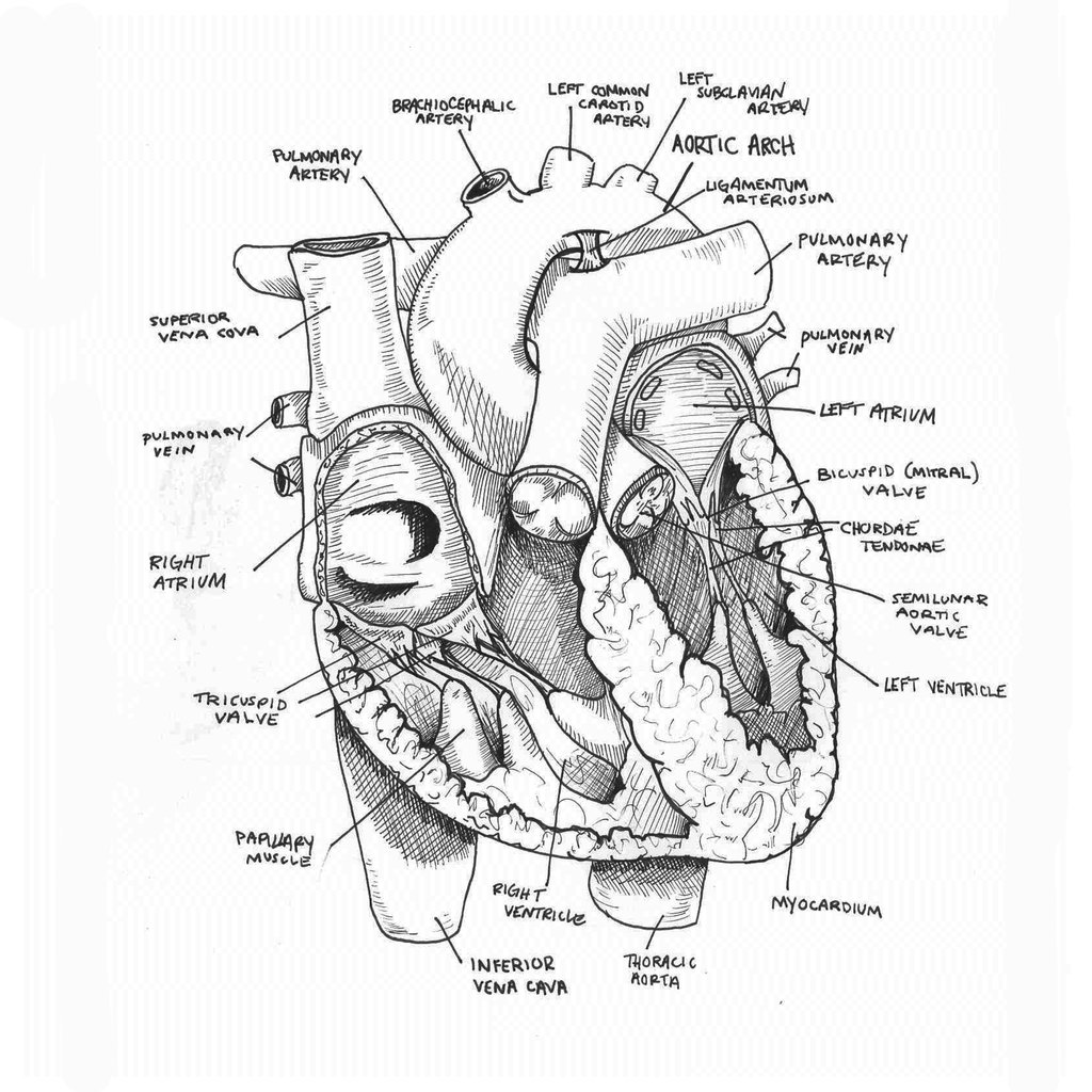
Heart Drawing Biology - This worksheet is designed to help a level students perfect their biological drawing technique. The heart is a muscular organ that pumps blood throughout the body. Web this post will focus on how i teach the structure of the heart so pupils can identify the four chambers of the heart, the vessels of the heart, which parts of the heart contain oxygenated or deoxygenated blood, and finally the pupils should be able to describe the route blood takes through the heart. So, grab your pencil and paper, and let’s get. Base (posterior), diaphragmatic (inferior), sternocostal (anterior), and left and right pulmonary surfaces. Plus, you may just learn something new along the way. The heart is a hollow, muscular organ located in the chest cavity. This video will help you to draw internal structure of human heart ( ಮಾನವ ಹೃದಯ. June 20, 2023 fact checked. The heart is a hollow, muscular organ located in the chest cavity. Plus, you may just learn something new along the way. After students finish their heart drawing they will trace the path of blood through the heart by adding arrows to show how blood flow from the heart, pulmonary circulation and systemic circulation. This is a quick way to learn how to draw the heart and some of the associated. The. How to draw the internal structure of the heart. So, grab your pencil and paper, and let’s get. Blood flow through the heart. Web the circulatory system (ccea) the heart blood is pumped away from the heart at high pressure in arteries, and returns to the heart at low pressure in veins. This is a quick way to learn how. Web dr matt & dr mike. Drawing a human heart is easier than you may think. The university of waikato te whare wānanga o waikato published 16 june 2017 referencing hub media. After reading this article you will learn about the structure of human heart. The blue lines in the drawing indicate the path of transmission of electrical signals through. Web your heart sure does work hard, but that doesn’t mean you have to work hard to draw it! This video will help you to draw internal structure of human heart ( ಮಾನವ ಹೃದಯ. So, grab your pencil and paper, and let’s get. Great vessels of the heart. This worksheet is designed to help a level students perfect their biological. This is a quick way to learn how to draw the heart and some of the associated. Even if you have never taught the heart before, do not worry. The human heart has a mass of around 300g and is roughly the size of a closed fist. Make a long cut down through the aorta and the left ventricle to. The heart is a muscular organ that pumps blood throughout the body. Great vessels of the heart. The heart is a hollow, muscular organ located in the chest cavity. So, grab your pencil and paper, and let’s get. This will also help you to draw the structure and diagram of human heart. The heart is one of the most important organs in a living being’s body, as it helps to keep blood flowing and ultimately helps to keep us alive! Web 1 minute heart. The human heart has a mass of around 300g and is roughly the size of a closed fist. It is located in the middle cavity of the chest,. In this drawing of the heart, the numbers refer to (1) the sinoatrial node and (2) the atrioventricular node. Even if you have never taught the heart before, do not worry. After reading this article you will learn about the structure of human heart. Web drawing an anatomical heart may seem like a complex task, but with the right approach,. Blood flow through the heart. Web about press copyright contact us creators advertise developers terms privacy policy & safety how youtube works test new features nfl sunday ticket press copyright. Great vessels of the heart. Web drawing an anatomical heart may seem like a complex task, but with the right approach, it can be easy and fun. 41k views 1. So, grab your pencil and paper, and let’s get. Web heart, organ that serves as a pump to circulate the blood. This will also help you to draw the structure and diagram of human heart. Web this post will focus on how i teach the structure of the heart so pupils can identify the four chambers of the heart, the. June 20, 2023 fact checked. The heart is one of the most important organs in a living being’s body, as it helps to keep blood flowing and ultimately helps to keep us alive! After reading this article you will learn about the structure of human heart. In this interactive, you can label parts of the human heart. Blood flow through the heart. So, grab your pencil and paper, and let’s get. Web this post will focus on how i teach the structure of the heart so pupils can identify the four chambers of the heart, the vessels of the heart, which parts of the heart contain oxygenated or deoxygenated blood, and finally the pupils should be able to describe the route blood takes through the heart. The human heart has a mass of around 300g and is roughly the size of a closed fist. The left and right side of the heart is separated by a muscular wall, the septum. It consists of four chambers and associated blood vessels. It is protected in the chest cavity by the pericardium, a tough and fibrous sac. This is a quick way to learn how to draw the heart and some of the associated. Base (posterior), diaphragmatic (inferior), sternocostal (anterior), and left and right pulmonary surfaces. Web your heart sure does work hard, but that doesn’t mean you have to work hard to draw it! In most people, the heart is located on the left side of the chest, beneath the breastbone. Web k examine the surface of the heart for blood vessels.
Human Heart Sketchbook study by bluesytealyren on DeviantArt Sketch

How to draw Heart Biology drawing for science students YouTube

How to draw Human Heart with colour Human Heart labelled diagram

The human heart Biology assignment YouTube

How To Draw Human Heart Diagram

human heart anatomy biology healthy image Stock Vector Image & Art

Anatomical Drawing Heart at GetDrawings Free download

Cardiac cycle and the Human Heart A* understanding for iGCSE Biology 2

how to draw human heart diagram easy/human heart drawing YouTube

How to Draw the Internal Structure of the Heart 14 Steps
This Video Will Help You To Draw Internal Structure Of Human Heart ( ಮಾನವ ಹೃದಯ.
41K Views 1 Year Ago Cardiovascular System.
The Heart Is A Hollow, Muscular Organ Located In The Chest Cavity.
Web Dr Matt & Dr Mike.
Related Post: