Drawing Of Stratified Squamous Epithelium
Drawing Of Stratified Squamous Epithelium - It is continuously replacing itself by division of the basal layer of cells. The apical cells appear squamous, whereas the basal layer contains either. The drawings of histology images were. Web if it is a stratified epithelium draw all the layers. Web stratified squamous epithelium occurs on numerous surfaces where it provides an important protective function. Web stratified squamous epithelium diagram and structure; It is made of four or five layers of epithelial cells, depending on its location in the body. First, the labeled diagram of. Medical school university of minnesota minneapolis, mn. Draw your structures proportionately to their size in your microscope’s field of view. A stratified squamous epithelium consists of squamous (flattened) epithelial cells arranged in layers upon a basal membrane. Use the image slider below to learn how to use a. The apical cells appear squamous, whereas the basal layer contains either. Web stratified squamous epithelium occurs on numerous surfaces where it provides an important protective function. Web keratinized stratified squamous epithelium is. Web here, i will provide both hand drawings and real microscope figures of stratified squamous epithelium (keratinized and nonkeratinized). The drawings of histology images were. Use the image slider below to learn how to use a. A stratified squamous epithelium has multiple layers of cells. Web stratified squamous epithelium diagram and structure; Web keratinized stratified squamous epithelium is a type of stratified epithelium that contains numerous layers of squamous cells, called keratinocytes, in which the superficial. 19k views 2 years ago cell biology. A stratified squamous epithelium has multiple layers of cells. Web stratified squamous epithelium occurs on numerous surfaces where it provides an important protective function. The apical cells appear squamous,. Web here, i will provide both hand drawings and real microscope figures of stratified squamous epithelium (keratinized and nonkeratinized). Web stratified squamous epithelium diagram and structure; 19k views 2 years ago cell biology. Web stratified squamous nonkeratinized epithelium. It is continuously replacing itself by division of the basal layer of cells. Web keratinized stratified squamous epithelium is a type of stratified epithelium that contains numerous layers of squamous cells, called keratinocytes, in which the superficial. The apical cells appear squamous, whereas the basal layer contains either. Web the epidermis is composed of keratinized, stratified squamous epithelium. Web structure of the stratified squamous epithelium. Fill in the blanks next to your drawing. Web simple squamous epithelia are nearly invisible when viewed from the apical surface due to their extreme thinness, so all images of simple squamous in this chapter will be cross. This epithelium protects against physical and chemical wear and tear. Web stratified epithelia consist of multiple layers of cells, with one layer anchored to the basement membrane, known as the. Web simple squamous epithelia are nearly invisible when viewed from the apical surface due to their extreme thinness, so all images of simple squamous in this chapter will be cross. Web stratified squamous epithelium occurs on numerous surfaces where it provides an important protective function. Stratified squamous epithelia are tissues formed from multiple layers of cells resting on a basement. Web simple squamous epithelia are nearly invisible when viewed from the apical surface due to their extreme thinness, so all images of simple squamous in this chapter will be cross. Web here, i will provide both hand drawings and real microscope figures of stratified squamous epithelium (keratinized and nonkeratinized). A stratified squamous epithelium has multiple layers of cells. First, the. Web keratinized stratified squamous epithelium is a type of stratified epithelium that contains numerous layers of squamous cells, called keratinocytes, in which the superficial. Medical school university of minnesota minneapolis, mn. 19k views 2 years ago cell biology. A stratified squamous epithelium consists of squamous (flattened) epithelial cells arranged in layers upon a basal membrane. Web stratified squamous nonkeratinized epithelium. The apical cells appear squamous, whereas the basal layer contains either. It is continuously replacing itself by division of the basal layer of cells. Use the image slider below to learn how to use a. Stratified squamous epithelia are tissues formed from multiple layers of cells resting on a basement membrane, with the. It is made of four or five. 4.5k views 2 years ago. First, the labeled diagram of. The apical cells appear squamous, whereas the basal layer contains either. Web stratified epithelia consist of multiple layers of cells, with one layer anchored to the basement membrane, known as the basal layer. Classification of stratified squamous epithelium. This epithelium protects against physical and chemical wear and tear. 19k views 2 years ago cell biology. The drawings of histology images were. It is made of four or five layers of epithelial cells, depending on its location in the body. Web stratified squamous epithelium diagram and structure; A stratified epithelium consists of several stacked layers of cells. Web the first pages illustrate introductory concepts for those new to microscopy as well as definitions of commonly used histology terms. Web stratified squamous epithelium definition. Web here, i will provide both hand drawings and real microscope figures of stratified squamous epithelium (keratinized and nonkeratinized). Web keratinized stratified squamous epithelium is a type of stratified epithelium that contains numerous layers of squamous cells, called keratinocytes, in which the superficial. Use the image slider below to learn how to use a.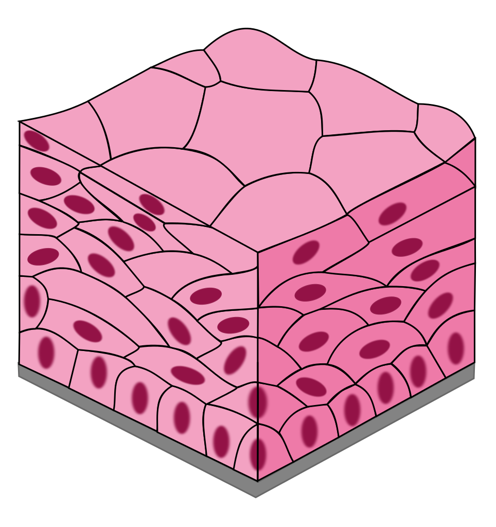
Epithelial tissues

Stratified squamous epithelium, illustration Stock Image F024/3098
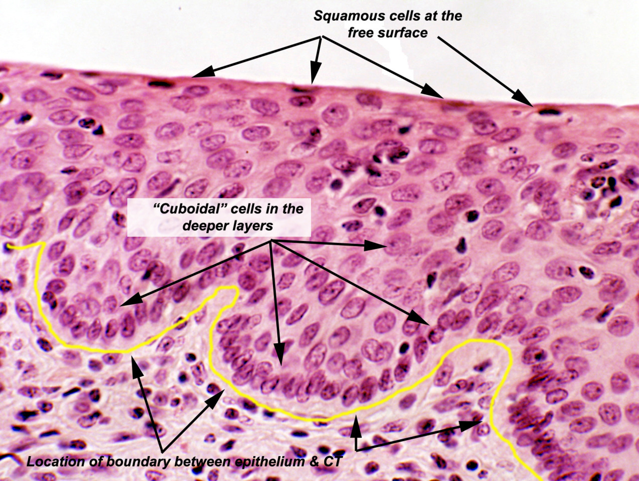
Epithelial Tissue Locations Stratified Squamous Epithelium Histology
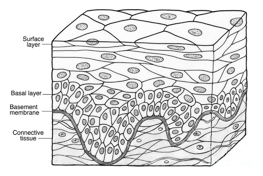
Illustration Of Stratified Squamous Photograph by Science Source Pixels
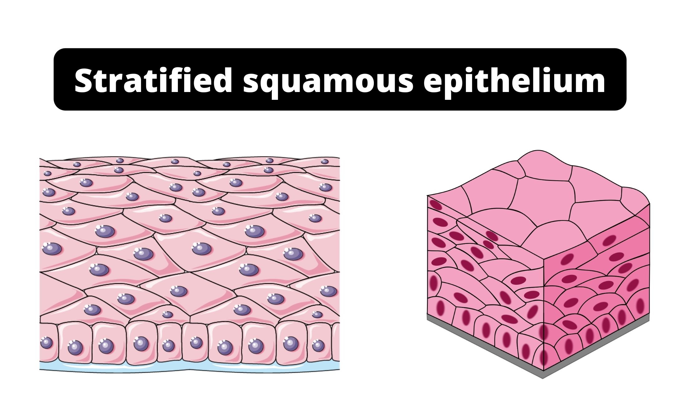
Stratified squamous epithelium Function, Definition, Location, Types.

Oral mucosa Lamina propria Wikipedia Basement membrane

Illustration of Stratified Squamous Epithelium Stock Photo Alamy

Stratified Squamous Epithelium Overview, Function & Location Lesson

How to draw stratified squamous epithelium easy way YouTube
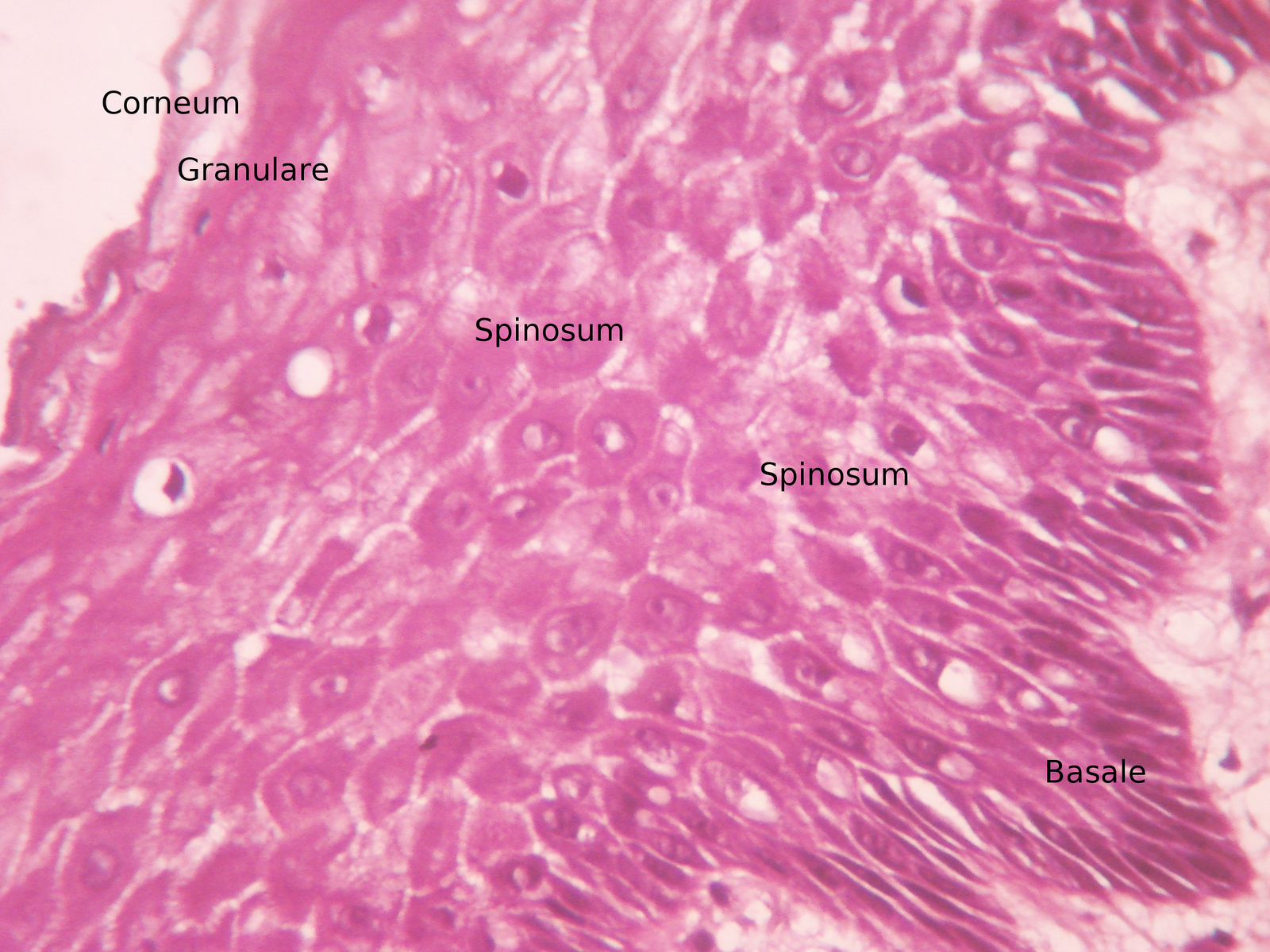
into the roots Stratified squamous epithelium
Web Simple Squamous Epithelia Are Nearly Invisible When Viewed From The Apical Surface Due To Their Extreme Thinness, So All Images Of Simple Squamous In This Chapter Will Be Cross.
Web Stratified Squamous Epithelium Is The Most Common Type Of Stratified Epithelium In The Human Body.
Medical School University Of Minnesota Minneapolis, Mn.
Draw Your Structures Proportionately To Their Size In Your Microscope’s Field Of View.
Related Post: