Drawing Of A Long Bone
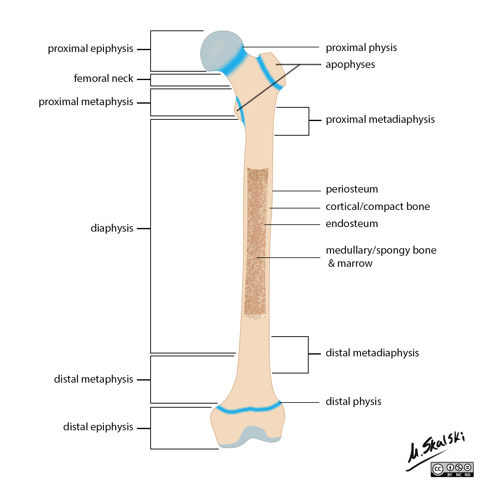
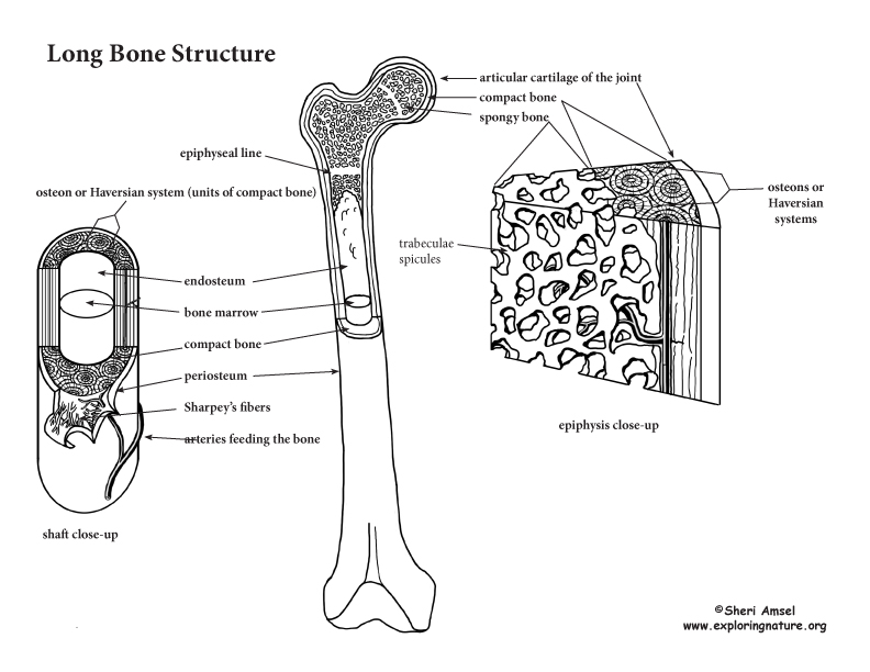
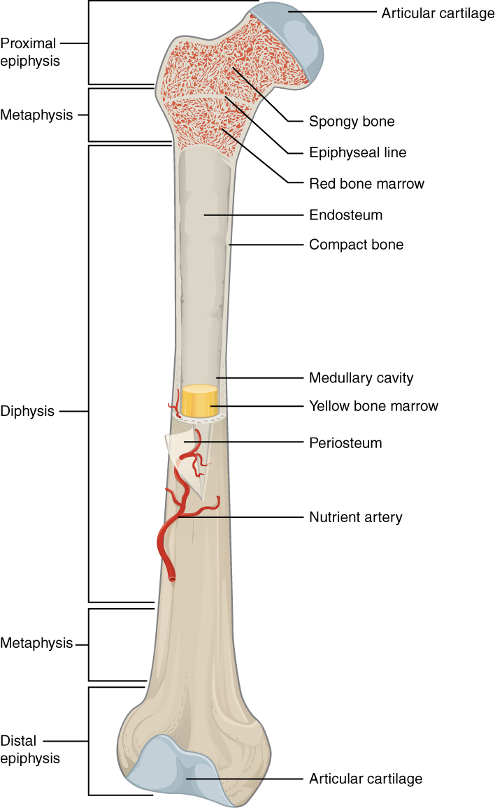
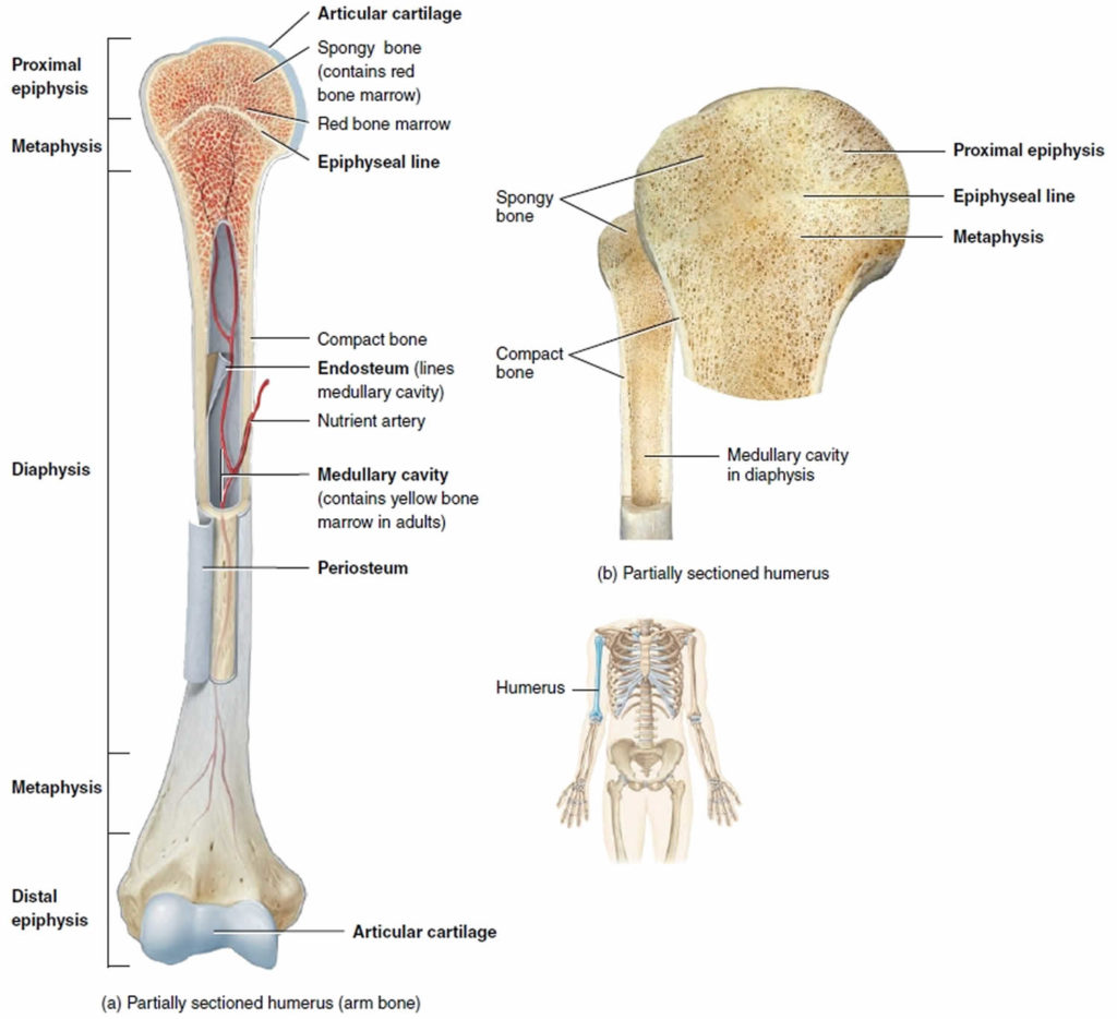
Drawing Of A Long Bone - Web a long bone has two main regions: J = epiphyseal line (growth plate) coloring worksheet for this image. Define and list examples of bone markings. The hollow region in the diaphysis is called the medullary cavity, which is. Identify the anatomical features of a bone. The diaphysis is the tubular shaft that runs between the proximal and distal ends of the bone. Learn about the bones, joints, and skeletal anatomy of the human body. The diaphysis and the epiphysis. Their structure & parts (shaft/diaphysis, epiphyses, metaphysis) with labeled diagram. The diaphysis and the epiphysis. Web c = articular cartilage. G = medullary cavity (yellow marrow) h = endosteum. New 3d rotate and zoom. Define and list examples of bone markings. A long bone has two parts: New 3d rotate and zoom. This article will provide detailed information on a long bone’s gross and microscopic features with a labeled diagram. The structure of a long bone allows for the best visualization of all of the parts of a bone (figure 1). Web gross anatomy of bone. G = medullary cavity (yellow marrow) h = endosteum. Long bones, such as the femur, are longer than they are wide. The human body is a complex, amazing biological machine. J = epiphyseal line (growth plate) coloring worksheet for this image. The diaphysis and the epiphysis. The diaphysis is the tubular shaft that runs between the proximal and distal ends of the bone. The diaphysis and the epiphysis. Web in this video i will be show you structure of long bone diagram draw the diagrams of science and get💯 marks. The diaphysis is the tubular shaft that runs between the proximal and distal ends of the bone. Learn about the bones, joints, and skeletal anatomy of the human body. The long bones are. The diaphysis is the hollow, tubular shaft that runs between the proximal and distal ends of the bone. G = medullary cavity (yellow marrow) h = endosteum. Anatomy of a long bone. Web a long bone has two main regions: Their structure & parts (shaft/diaphysis, epiphyses, metaphysis) with labeled diagram. Web a long bone has two main regions: The diaphysis is the tubular shaft that runs between the proximal and distal ends of the bone. Flat bones are thin, but are often curved, such as the ribs. How to draw humerus bone/humerus bone drawing it is very easy drawing detailed. G = medullary cavity (yellow marrow) h = endosteum. Explore the skeletal system with our interactive 3d anatomy models. J = epiphyseal line (growth plate) coloring worksheet for this image. Web anatomy of a long bone. Identify the anatomical features of a bone. Web definition of long bones in the body, with a list of names. The epiphysial plate has been closed in this bone and has become the epiphyseal line after puberty. The femur) consists of epiphyses, metaphyses and a diaphysis (shaft). Case courtesy of dr matt skalski, radiopaedia.org. Epiphysis), the bulky ends on both sides. The diaphysis and the epiphysis. A typical long bone shows the gross anatomical characteristics of bone. Epiphysis), the bulky ends on both sides. The femur) consists of epiphyses, metaphyses and a diaphysis (shaft). Long bones, especially the femur and tibia, are subjected to most of the load during daily activities and they are crucial for skeletal mobility. The diaphysis and the epiphysis. There is a narrow section called (3) metaphysis between the diaphysis and epiphysis. The diaphysis and the epiphysis. Web the structure of a long bone allows for the best visualization of all of the parts of a bone (figure 5.3.1 5.3. The diaphysis is the narrow, tubular shaft that runs between the two bulbous ends of the bone. The diaphysis. The diaphysis and the epiphysis ( figure 6.3.1). Web c = articular cartilage. The diaphysis is the hollow, tubular shaft that runs between the proximal and distal ends of the bone. Web a long bone has two main regions: Explore basic bone structure, upper & lower body, hands, feet, details, and shading. Web anatomy of a long bone. Web mishu drawing academy. The human body is a complex, amazing biological machine. They are one of five types of bones: The diaphysis and the epiphysis. Web in this video i will be show you structure of long bone diagram draw the diagrams of science and get💯 marks. The epiphysial plate has been closed in this bone and has become the epiphyseal line after puberty. Web the structure of a long bone allows for the best visualization of all of the parts of a bone. Learn about the bones, joints, and skeletal anatomy of the human body. 232k views 5 years ago introduction to anatomy and physiology. Long, short, flat, irregular and sesamoid.
Radiopaedia Drawing Anatomy of long bones (femur) English labels

Draw a diagram of a long bone and label the structures, iden Quizlet

Bones Anatomy of Long Bones

Long Bone Anatomy Drawn & Defined YouTube

Anatomy Of The Long Bone

Bone Cross Section Diagram Labeled / Long Bone High Res Stock Images

Long bone Wikipedia

Long Bone Diagram And Labelling vrogue.co

Long bone anatomy, structure, parts, function and fracture types

Sketch a typical long bone, and label its epiphyses, diaphys Quizlet
In This Video We Discuss The Parts Of A Long Bone And Some Of The Functions Of Each Of Those Bone Parts.
Case Courtesy Of Dr Matt Skalski, Radiopaedia.org.
Identify The Anatomical Features Of A Bone.
New 3D Rotate And Zoom.
Related Post: