Draw The F As Seen In The Low Power Field
Draw The F As Seen In The Low Power Field - You have been a ked to prepare slide with the letter fon it (as shown below). This may be sufficient to view your chosen organism. Scanning lens (4x) _____mm, _____ μm. Look at the objectives of your microscope and record the magnification power. Web in the circle below. You have been asked to prepare a slide with the letter fon it (as shown below), in the. Web notice there is a danger of smashing the objective lens into the slide if you were to use the coarse focus. Web one would like to view at low power, and center it in the field of view. Low power lens (10x) _____mm, _____ μm. Web describe the proper procedure for preparing a wet mount. Web you have been asked to prepare a slide with the letter f on it (as shown below). What happens to the “e” as you move the slide to the right? Web calculate the field size in micrometers (μm). Then carefully position the coverslip. Web draw the organisms that you see. Last, slowly and carefully lower. What happens to the “e” as you move the slide to the right? Web notice there is a danger of smashing the objective lens into the slide if you were to use the coarse focus. Web calculate the field size in micrometers (μm). Then carefully position the coverslip. Also indicate the estimated cell size in micrometers under your drawing. If high power is 10x more magnification than the low power, the field of view will be 1/10 as big. Anatomy and physiology questions and answers. You should see other millimeter markings in your field of view. Web center the specimen in microscope field by moving the stage. This problem has been solved!. Bring the letter into focus under low power using the procedures described above. Web describe the proper procedure for preparing a wet mount. Then carefully position the coverslip. You have been asked to prepare a slide with the letter fon it (as shown below), in the. Bring the letter into focus under low power using the procedures described above. You should see other millimeter markings in your field of view. Estimate the length (longest dimension) of the object in urn: Web in the circle below. 1 mm = 1000 μm. Web switch to low power (10x). This may be sufficient to view your chosen organism. Web you have been asked to prepare a slide with the letter k on it. Web calculate the field size in micrometers (μm). Describe the position of the “e.” 3. Draw the letter “e” as it appears under the microscope. If high power is 10x more magnification than the low power, the field of view will be 1/10 as big. Estimate the length (longest dimension) of the object in urn: Also indicate the estimated cell size in micrometers under your drawing. You have been a ked to prepare slide with. This problem has been solved!. You have been asked to prepare a slide with the letter fon it (as shown below), in the. Web place one millimeter marking of your ruler at the far left hand side of the scanning power field. Also indicate the estimated cell size in micrometers under your drawing. Draw the letter “e” as it appears. This may be sufficient to view your chosen organism. You have been asked to prepare a slide with the letter fon it (as shown below), in the. Web switch to low power (10x). Web draw the organisms that you see. Web you have been asked to prepare a slide with the letter k on it. Web switch to low power (10x). Estimate the length (longest dimension) of the object in urn: This may be sufficient to view your chosen organism. Web draw the organisms that you see. Total magnification = field diameter = 1.6 mm. Draw the letter “e” as it appears under the microscope. Bring the letter into focus under low power using the procedures described above. Anatomy and physiology questions and answers. Estimate the length (longest dimension) of the object in urn: Total magnification = field diameter = 1.6 mm. Web now center the slide of the letter “e” on the stage with the “e” in its normal upright position. Web in the circle below. Scanning lens (4x) _____mm, _____ μm. You have been asked to prepare a slide with the letter fon it (as shown below), in the. Web one would like to view at low power, and center it in the field of view. Web you have been asked to prepare a slide with the letter f f f on it (as shown below). Look at the objectives of your microscope and record the magnification power. Try to note how it moves and do your best to draw it as you see it, unless you need more. What happens to the “e” as you move the slide to the right? Ned mated diameter of mm 7. Web draw the organisms that you see.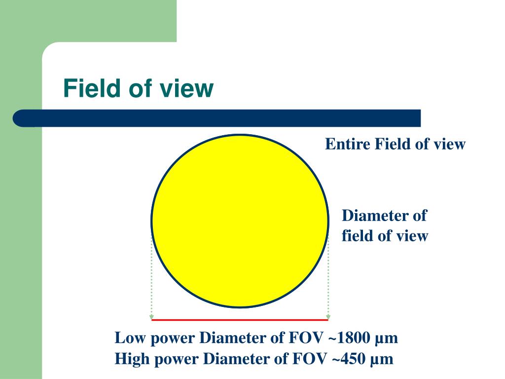
PPT Introduction to the Light Microscope PowerPoint Presentation

Pathological findings. Lowpower field (a) and highpower field (b) of

How to design progressive lenses Knowledgebase
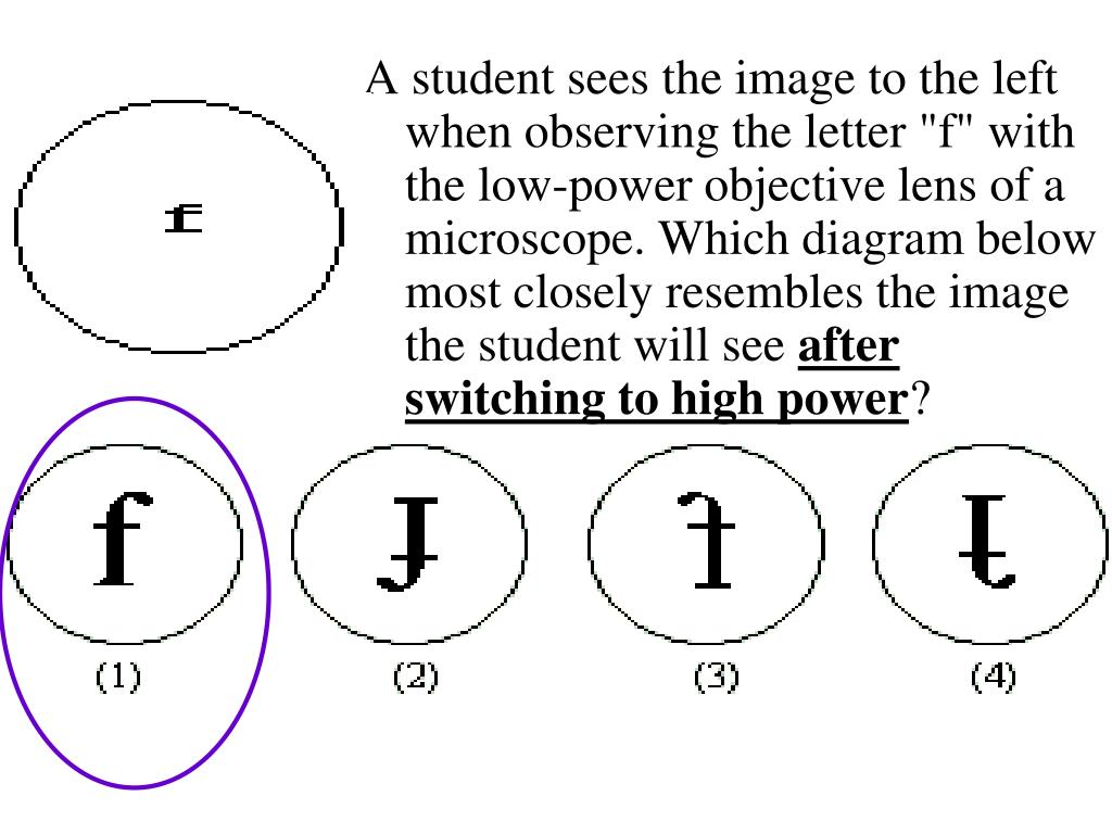
PPT Microscope Review PowerPoint Presentation, free download ID6690365
![[PDF] LowPower FieldProgrammable VLSI Using Multiple Supply Voltages](https://d3i71xaburhd42.cloudfront.net/a596f5f5e9bac8130e7a195ecfdb588100630026/6-Figure16-1.png)
[PDF] LowPower FieldProgrammable VLSI Using Multiple Supply Voltages
LUNAR Lowpower Field Procedures
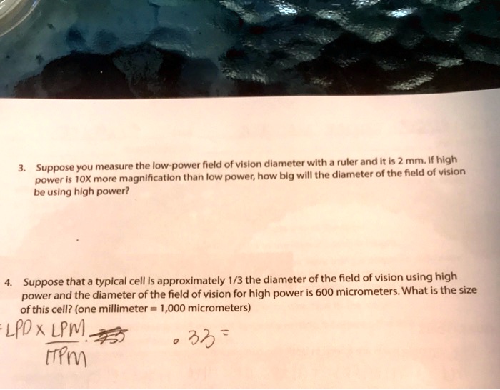
SOLVED Suppose you measure the low power field of vislon dlameter with
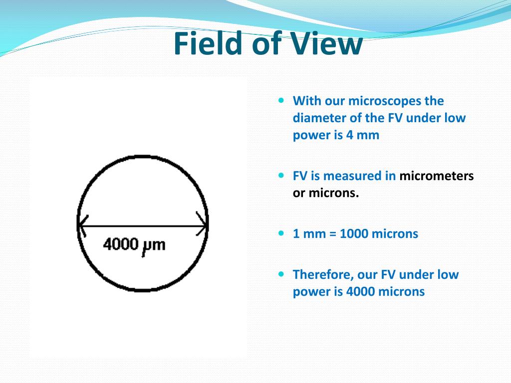
PPT The Microscope PowerPoint Presentation, free download ID6006599

Figure 10 from LowPower FieldProgrammable VLSI Using Multiple Supply

How to Calculate the Low Power Magnification of a Microscope Estrella
This May Be Sufficient To View Your Chosen Organism.
Web Describe The Proper Procedure For Preparing A Wet Mount.
Web Notice There Is A Danger Of Smashing The Objective Lens Into The Slide If You Were To Use The Coarse Focus.
Look At Microscope From The Side:
Related Post: