Draw Sister Chromatids
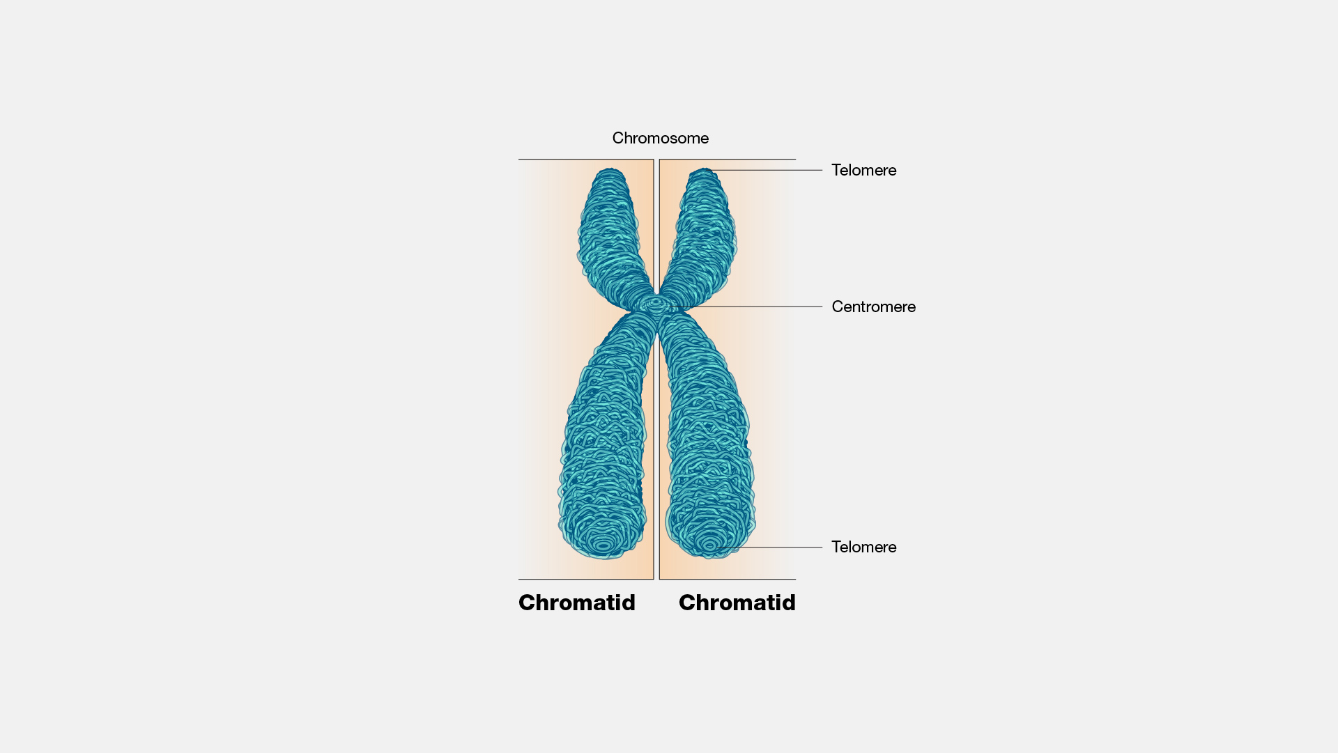
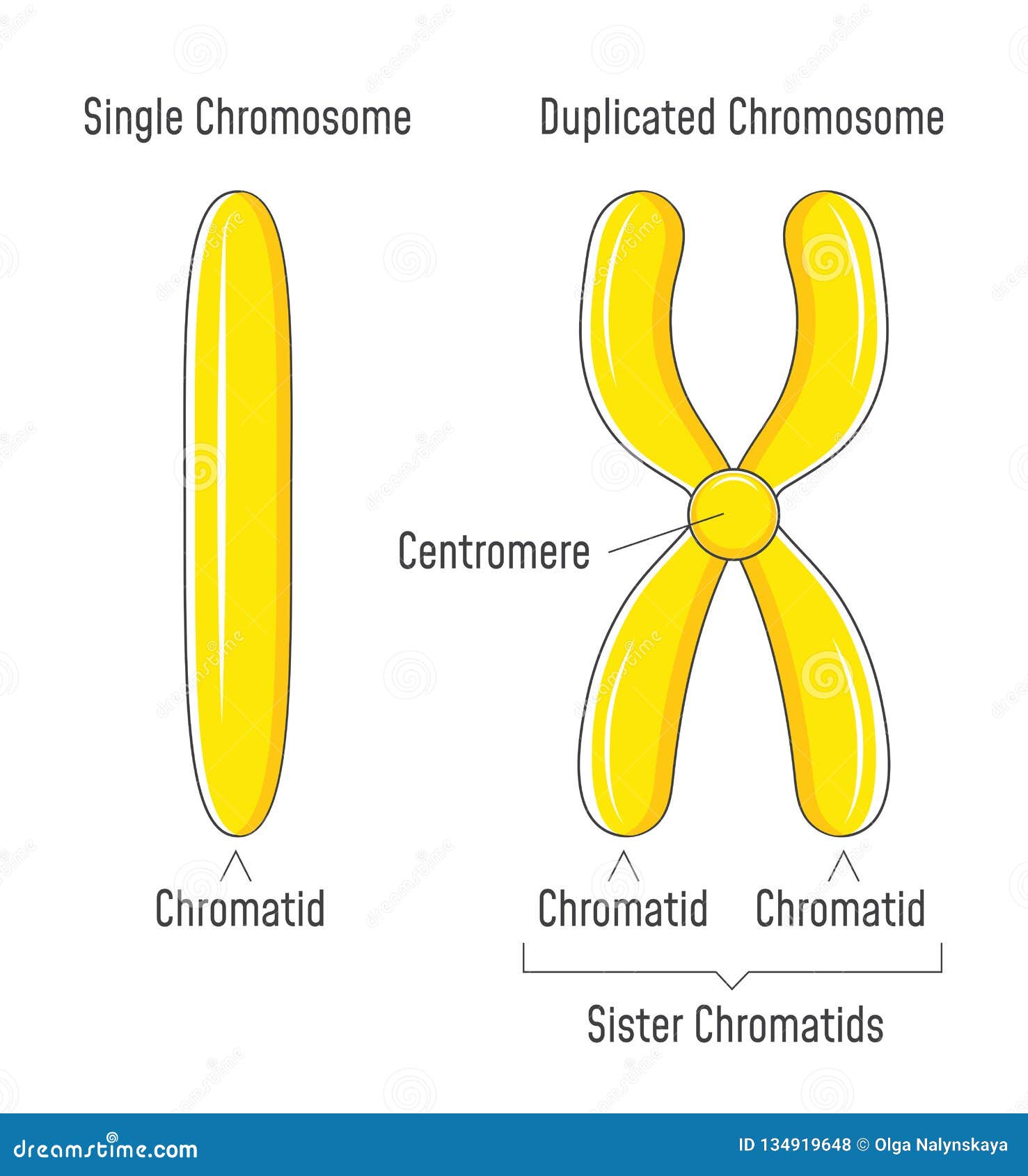
Draw Sister Chromatids - A kinetochore, a large, multimeric protein, joins the two strands at this section. And we can draw the blue chromosome. In meiosis i, cells go through four phases: During prophase i, chromosomes pair up and exchange genetic material, creating more variation. This is important for chromosomal separation during cell division. The sister chromatids are identical to one another and are attached to each other by proteins called cohesins. Crossing over occurs only during prophase i. The two sister chromatids are separated from each other into two different cells during mitosis or during the second division of meiosis. A chromatid is one of the two identical halves of a chromosome that has been replicated in preparation for cell division. Web chromosomes and cell division. Web eukaryotic chromosomes consist of two sister chromatids. Chromosome (label as duplicated or unduplicated), centromere, kinetochore, sister chromatids, nonsister chromatids, homologous pair (use a bracket when labeling), homolog (label each one), chiasma, sister chromatid cohesion, and gene loci, labeling the alleles of the f and h genes. Web as the two daughter dna strands are produced from the chromosomal dna. Although a cell needs to undergo interphase before entering meiosis, interphase is technically not part of meiosis. The two “sister” chromatids are joined at a constricted region of the chromosome called the centromere. Cohesin holds the chromatids together until anaphase ii. The mechanics of meiosis ii are similar to mitosis, except that each dividing cell has only one set of. Web sister chromatids definition. Cohesin holds the chromatids together until anaphase ii. Right now, both of these two sister chromatids combined are considered to be one chromosome. Chromosome replication takes place during interphase of the cell cycle. Even though before replication it was still considered, the magenta stuff was still considered to be one chromosome. Web the second meiotic division, where sister chromatids separate, is like mitosis. • dna is replicated before meiosis so that all chromosomes consist of two sister chromatids. The sister chromatids are identical to one another and are attached at a compressed region called the centromere. Web study with quizlet and memorize flashcards containing terms like draw a picture of a. Web a full set of sister chromatids is created during the synthesis ( s) phase of interphase, when all the chromosomes in a cell are replicated. Even though before replication it was still considered, the magenta stuff was still considered to be one chromosome. Web the second meiotic division, where sister chromatids separate, is like mitosis. And we can draw. Even though before replication it was still considered, the magenta stuff was still considered to be one chromosome. How do they differ?, what does diploid mean? Chromosome replication takes place during interphase of the cell cycle. In meiosis i, cells go through four phases: Although a cell needs to undergo interphase before entering meiosis, interphase is technically not part of. Although a cell needs to undergo interphase before entering meiosis, interphase is technically not part of meiosis. As a cell prepares to divide, it must make a copy of each of its chromosomes. Even though before replication it was still considered, the magenta stuff was still considered to be one chromosome. Web the sister chromatids separate from one another and. In meiosis i, cells go through four phases: Chromosome number stays the same when sister chromatids separate. Web eukaryotic chromosomes consist of two sister chromatids. Crossing over occurs only during prophase i. The two copies of a chromosome are called sister chromatids. Web two sister chromatids attach at a region called the centromere. Web the sister chromatids separate from one another and are pulled towards opposite poles of the cell. Cohesin holds the chromatids together until anaphase ii. In meiosis i, cells go through four phases: Although a cell needs to undergo interphase before entering meiosis, interphase is technically not part of. The sister chromatids are identical to one another and are attached to each other by proteins called cohesins. Web the second meiotic division, where sister chromatids separate, is like mitosis. Web two sister chromatids attach at a region called the centromere. Using the information above, compare these two simplified diagrams of mitosis and meiosis to visualize why cells are haploid. Using the information above, compare these two simplified diagrams of mitosis and meiosis to visualize why cells are haploid after meiosis i. This is important for chromosomal separation during cell division. Web a full set of sister chromatids is created during the synthesis ( s) phase of interphase, when all the chromosomes in a cell are replicated. Chromosome (label as duplicated or unduplicated), centromere, kinetochore, sister chromatids, nonsister chromatids, homologous pair (use a bracket when labeling), homolog (label each one), chiasma, sister chromatid cohesion, and gene loci, labeling the alleles of the f and h genes. And we can draw the blue chromosome. Sister chromatids and homologous chromosomes during interphase of the cell cycle, the dna is replicated. During cell division, they are separated from each other, and each daughter cell receives one copy of the chromosome. Cohesin holds the chromatids together until anaphase ii. Web cohesin forms rings that hold the sister chromatids together, whereas condensin forms rings that coil the chromosomes into highly compact forms. Sister chromatids are genetically identical copies or replicas of a single chromosome. The mitotic spindle also begins to develop. The two sister chromatids are separated from each other into two different cells during mitosis or during the second division of meiosis. Web during meiosis ii, the sister chromatids within the two daughter cells separate, forming four new haploid gametes. These copies remain attached until sister chromatids are separated or detached during cell division. Web during mitosis, the two sister chromatids that make up each chromosome separate from each other and move to opposite poles of the cell. Web the sister chromatids separate from one another and are pulled towards opposite poles of the cell.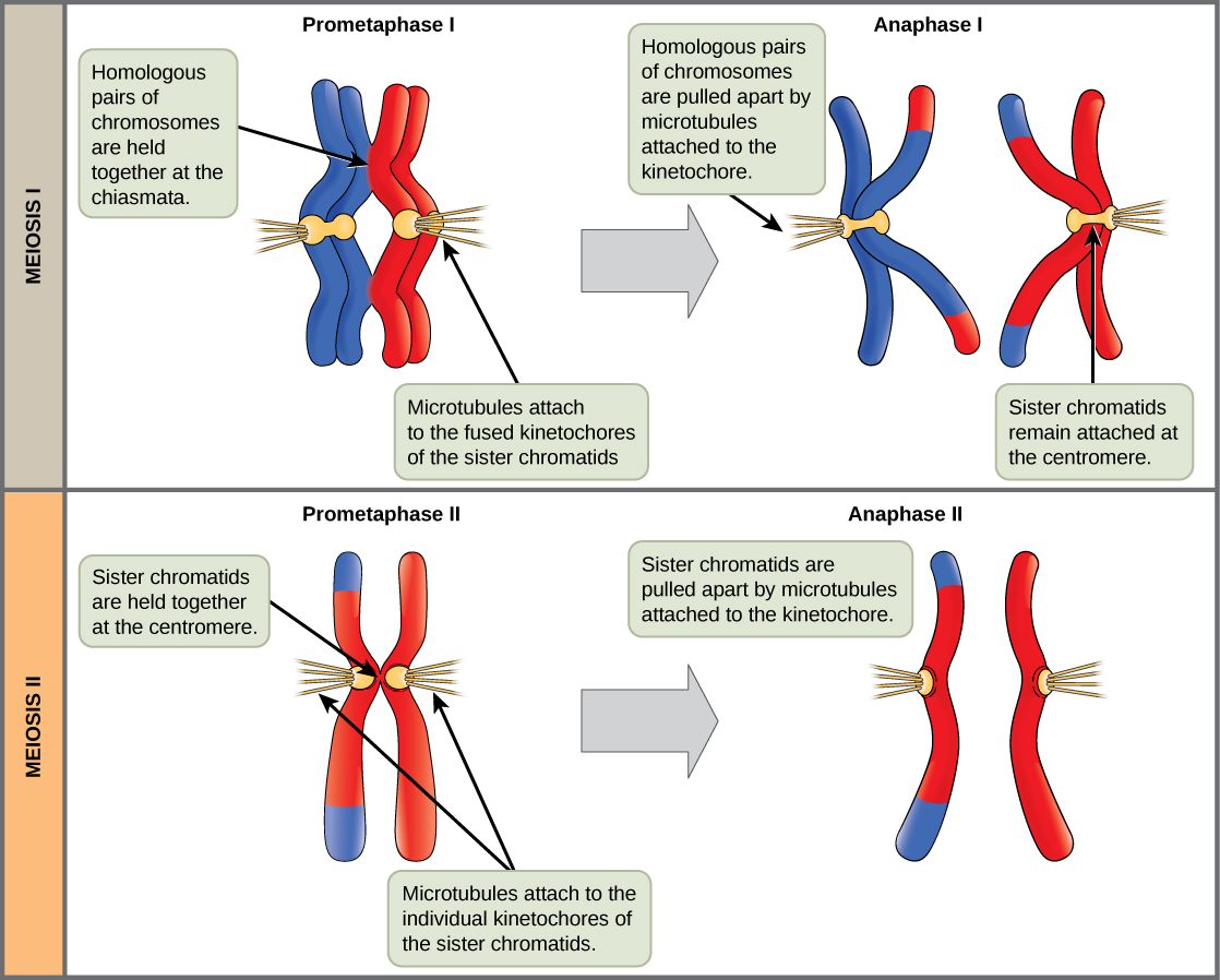
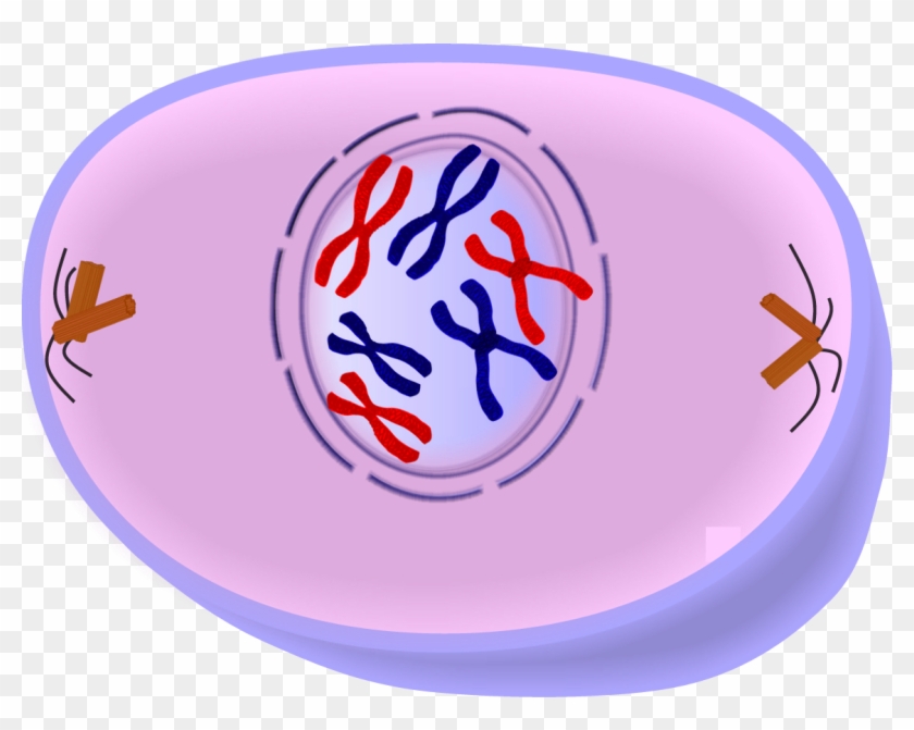
Meiosis II Biology for Majors I
Download Chromatin Drawing Chromosome Sister Chromatids Cell In
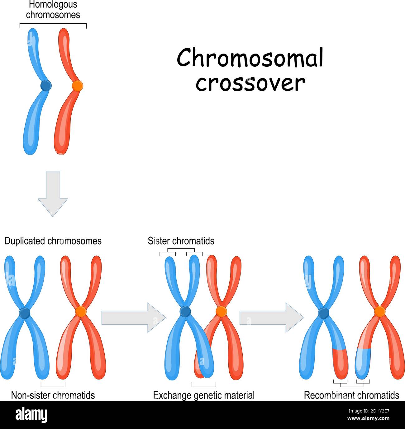
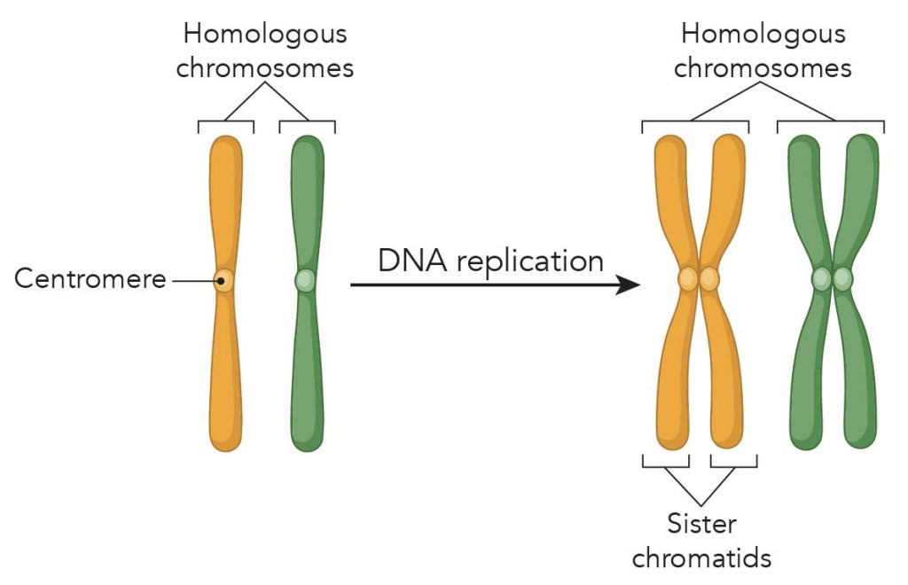
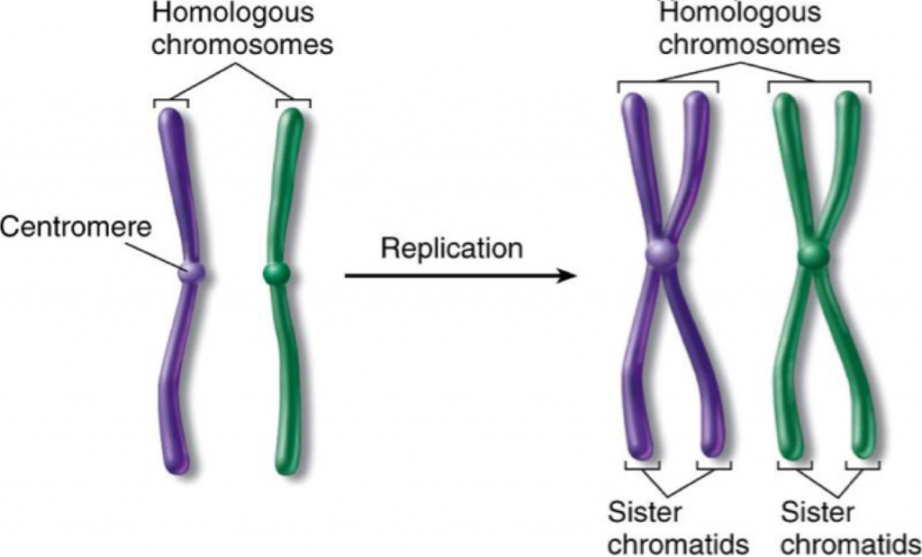
Homologous Chromosomes And Sister Chromatids
Centromere Definition and Examples Biology Online Dictionary
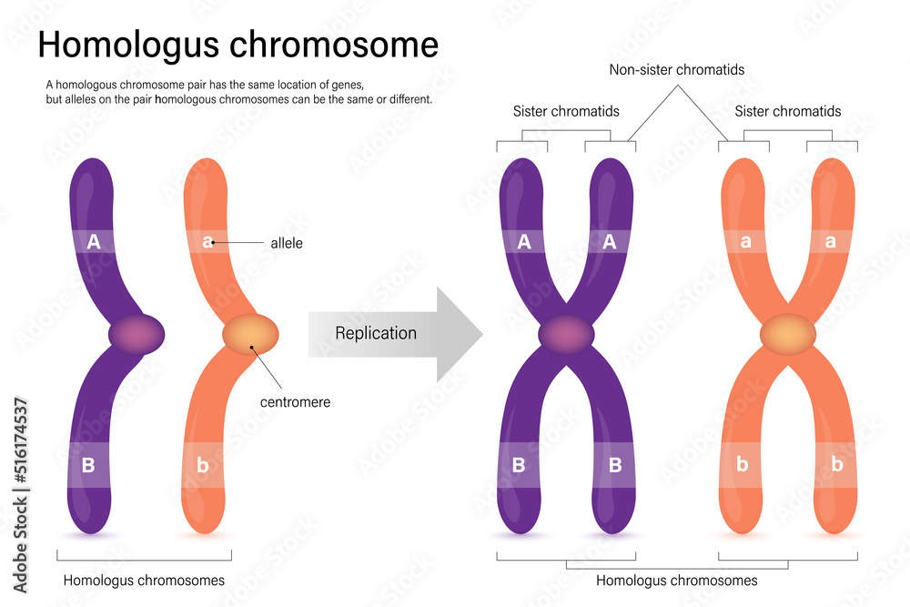
Diagram of homologus chromosome. Sister chromatids and Nonsister
What is Mitosis (Food model of mitosis) Rs' Science
3.2 Chromosomes The Biology Classroom
Homologous Chromosomes Definition, Functions & Examples
Unduplicated and Duplicated Chromosomes. Sister Chromatids Stock Vector

13.1 DNA Biology LibreTexts
Even Though Before Replication It Was Still Considered, The Magenta Stuff Was Still Considered To Be One Chromosome.
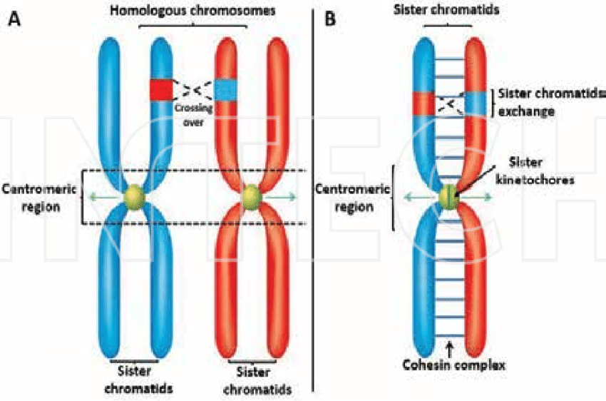
A Kinetochore, A Large, Multimeric Protein, Joins The Two Strands At This Section.
Web And It Has The Centromere That Connects These Two Sister Chromatids.
Web The Second Meiotic Division, Where Sister Chromatids Separate, Is Like Mitosis.
Related Post: