Draw And Label The Heart
Draw And Label The Heart - Web to draw the internal structure of the heart, start by sketching the 2 pulmonary veins to the lower left of the aorta and the bottom of the inferior vena cava slightly to the right of that. After all, we know that stress is bad for the heart! 14 views 1 year ago. The two atria and ventricles are separated from each other by a muscle wall called ‘septum’. Blood flow through the heart, cardiac circulation pathway, and anatomy of the heart. Drag and drop the text labels onto the boxes next to the diagram. Relate the structure of the heart to its function as a pump. Dissect a pig’s or sheep’s heart and label the main chambers, valves, vessels, and other structures. Compare systemic circulation to pulmonary circulation. Web medically reviewed by the healthline medical network — by the healthline editorial team — updated on january 20, 2018. Dr matt & dr mike. The heart is responsible for the circulation of blood in our body. Great vessels of the heart. The right side of the heart receives deoxygenated blood from the systemic veins and pumps it to the lungs for. Web cardiovascular heart diagram: Web the human heart, comprises four chambers: Selecting or hovering over a box will highlight each area in the diagram. The two upper chambers are called the left and the right atria, and the two lower chambers are known as the left and the right ventricles. Public domain license) learning objectives. The right side of the heart receives deoxygenated blood. After all, we know that stress is bad for the heart! Identify and trace the path of blood flow through the heart using the dissected heart. The upper two chambers of the heart are called auricles. Web function and anatomy of the heart made easy using labeled diagrams of cardiac structures and blood flow through the atria, ventricles, valves, aorta,. The heart is responsible for the circulation of blood in our body. Muscle and tissue make up this powerhouse organ. Web in this interactive, you can label parts of the human heart. Anatomical illustrations and structures, 3d model and photographs of dissection. Web best way to draw and label the heart! The heart is a hollow, muscular organ that pumps oxygenated blood throughout the body and deoxygenated blood to the lungs. Dissect a pig’s or sheep’s heart and label the main chambers, valves, vessels, and other structures. Selecting or hovering over a box will highlight each area in the diagram. Web in this interactive, you can label parts of the human. Identify and trace the path of blood flow through the heart using the dissected heart. Web in just a few minutes, you will be able to label the entire diagram shown below! Web the heart is located in the thoracic cavity medial to the lungs and posterior to the sternum. Blood flow through the heart, cardiac circulation pathway, and anatomy. 41k views 1 year ago cardiovascular system. Web the heart is located in the thoracic cavity medial to the lungs and posterior to the sternum. Web a well labeled human heart diagram given in this article will help you to understand its parts and functions. Web best way to draw and label the heart! Your heart sure does work hard,. Base (posterior), diaphragmatic (inferior), sternocostal (anterior), and left and right pulmonary surfaces. 41k views 1 year ago cardiovascular system. Rotate the 3d model to see how the heart's valves control blood flow between heart chambers and blood flow out of the heart. Drawing a human heart is easier than you may think. Great vessels of the heart. Web cardiovascular heart diagram: Web this interactive atlas of human heart anatomy is based on medical illustrations and cadaver photography. The inferior tip of the heart, known as the apex, rests just superior to the diaphragm. Compare systemic circulation to pulmonary circulation. The right side of the heart receives deoxygenated blood from the systemic veins and pumps it to the. Identify the veins and arteries of the coronary circulation system. Web cardiovascular heart diagram: Relate the structure of the heart to its function as a pump. In coordination with valves, the chambers work to keep blood flowing. The user can show or hide the anatomical labels which provide a useful tool to create illustrations perfectly adapted for teaching. Web the heart is located in the thoracic cavity medial to the lungs and posterior to the sternum. Compare systemic circulation to pulmonary circulation. The video above also provides an animation at the end to quiz yourself and test your knowledge! The inferior tip of the heart, known as the apex, rests just superior to the diaphragm. The heart is a hollow, muscular organ that pumps oxygenated blood throughout the body and deoxygenated blood to the lungs. Web best way to draw and label the heart! Base (posterior), diaphragmatic (inferior), sternocostal (anterior), and left and right pulmonary surfaces. 14 views 1 year ago. It’s your circulatory system ’s main organ. Do you want a fun way to learn the structure of the heart? Web a well labeled human heart diagram given in this article will help you to understand its parts and functions. The two upper chambers are called the left and the right atria, and the two lower chambers are known as the left and the right ventricles. Web this interactive atlas of human heart anatomy is based on medical illustrations and cadaver photography. Web the diagram of heart is beneficial for class 10 and 12 and is frequently asked in the examinations. Selecting or hovering over a box will highlight each area in the diagram. After all, we know that stress is bad for the heart!
How to Draw the Internal Structure of the Heart 14 Steps

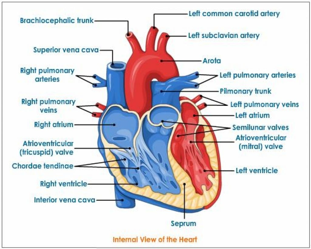
External Structure Of Heart Anatomy Diagram
:max_bytes(150000):strip_icc()/heart_exterior_anatomy-577d5cc23df78cb62c942f06.jpg)
The Anatomy of the Heart, Its Structures, and Functions
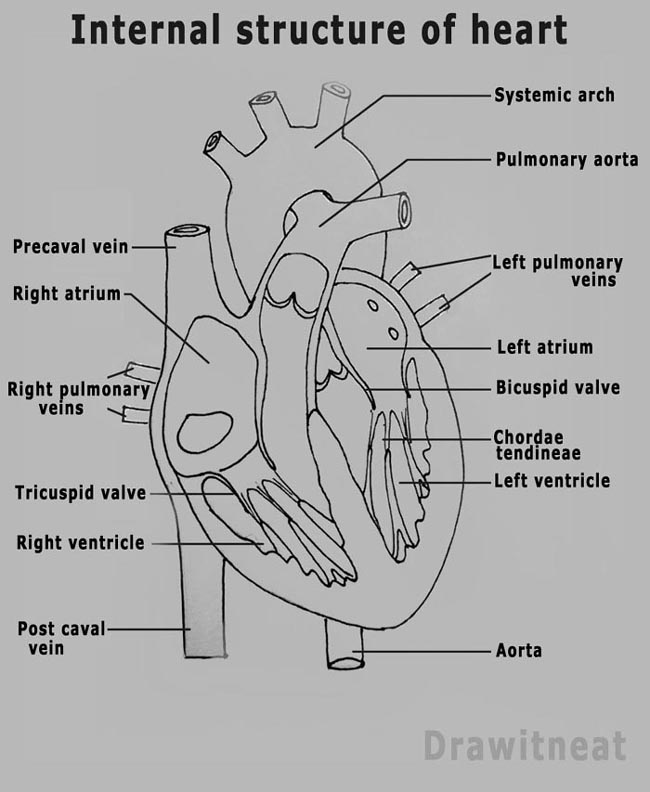
DRAW IT NEAT How to draw human heart labeled
Heart And Labels Drawing at GetDrawings Free download

How to Draw the Internal Structure of the Heart 13 Steps

humanheartdiagram Tim's Printables

FileHeart diagramen.svg Wikipedia
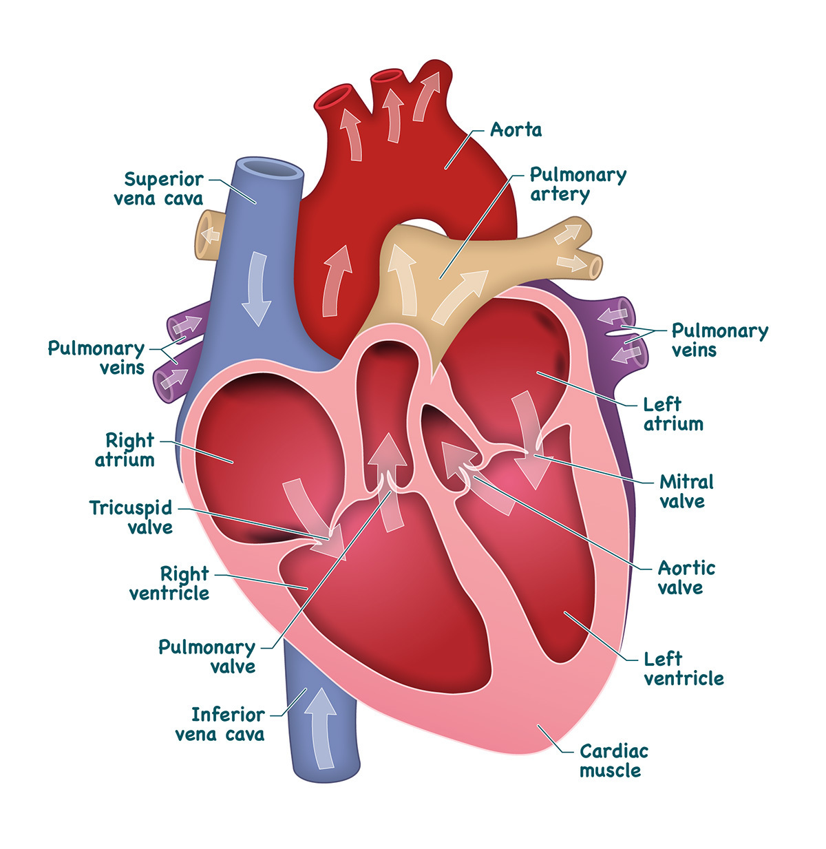
Heart Anatomy Labeled Diagram, Structures, Blood Flow, Function of
Heart And Labels Drawing at GetDrawings Free download
Web Identify The Tissue Layers Of The Heart.
The Two Atria And Ventricles Are Separated From Each Other By A Muscle Wall Called ‘Septum’.
Your Heart Contains Four Muscular Sections ( Chambers) That Briefly Hold Blood Before Moving It.
41K Views 1 Year Ago Cardiovascular System.
Related Post:
