Compact Bone Drawing
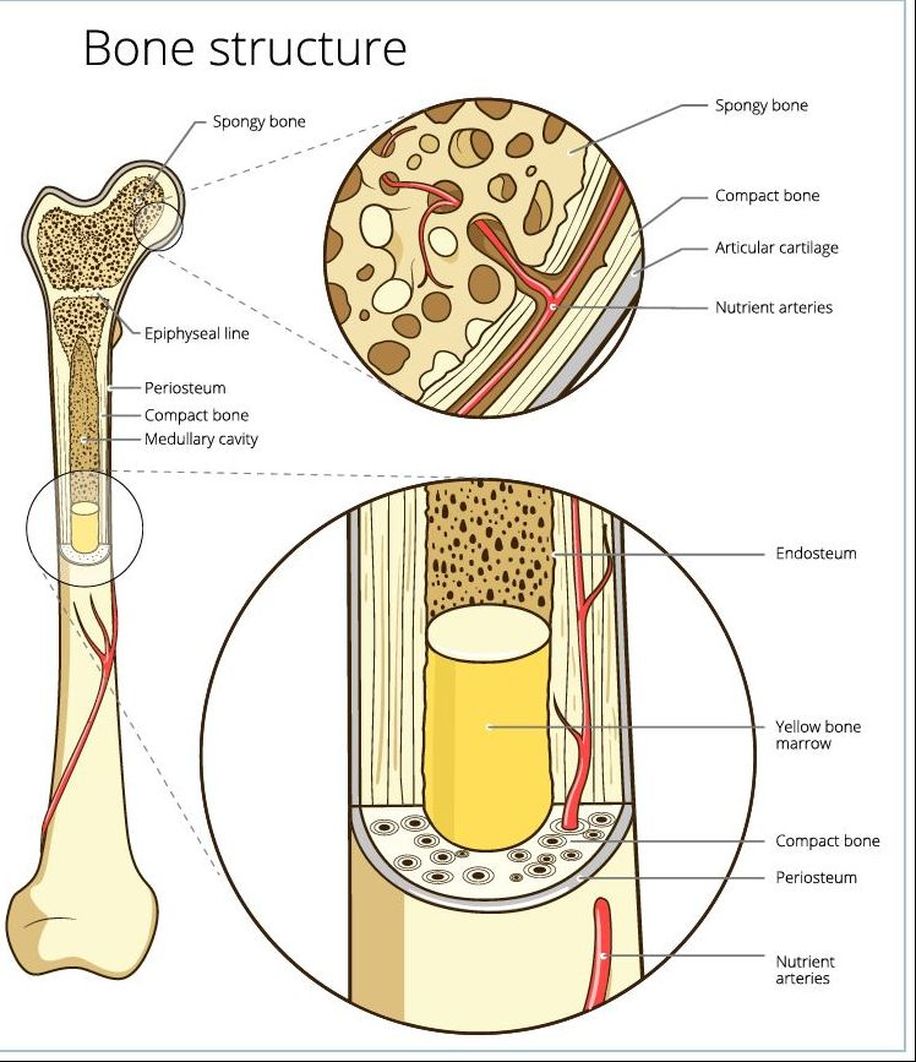
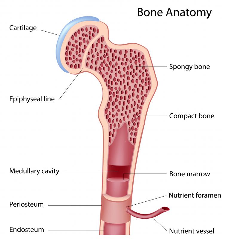
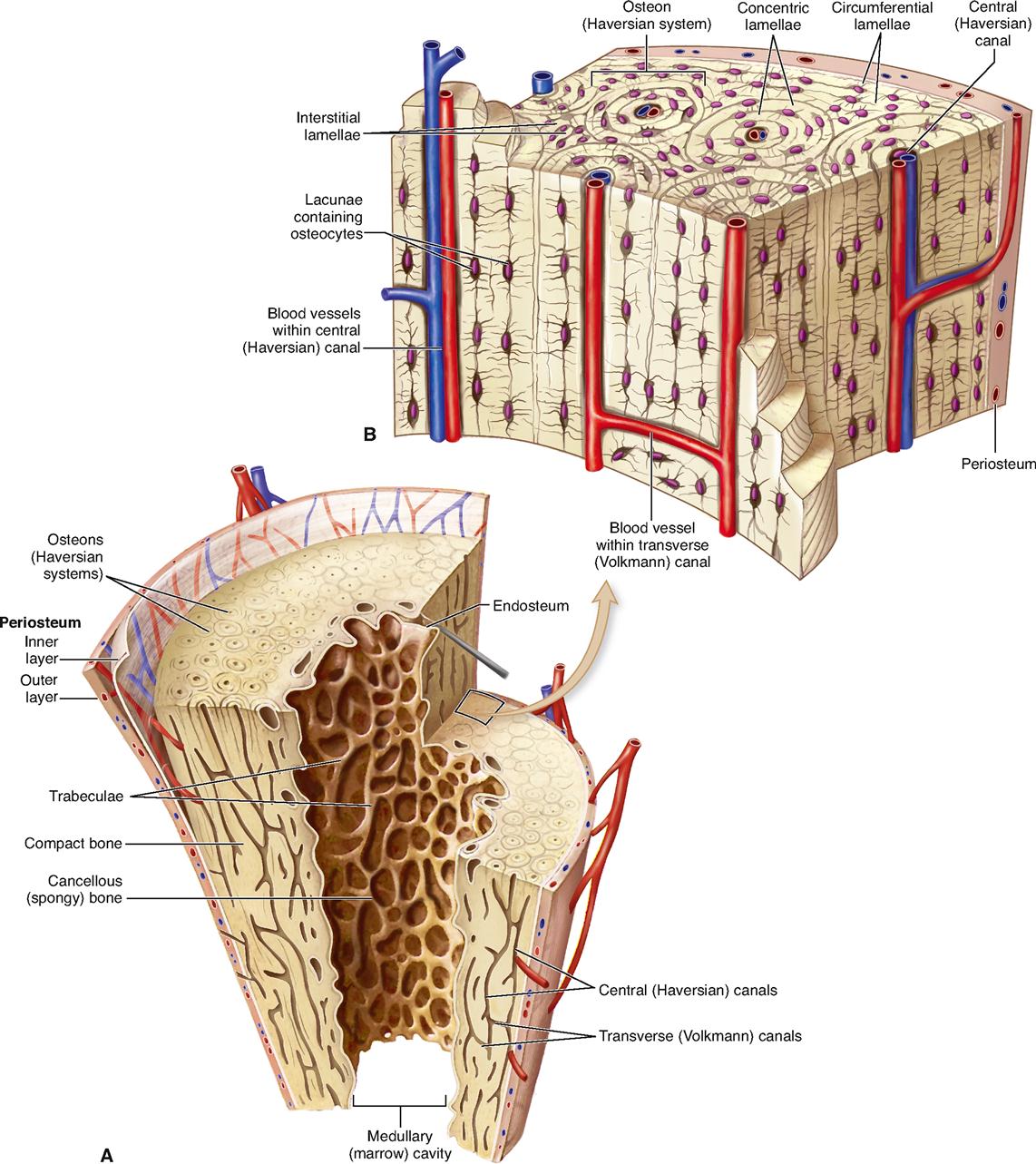
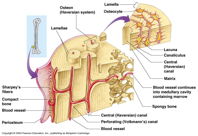
Compact Bone Drawing - Use the drawing tools tab to change the formatting of the pull quote text. The two layers of compact bone and the interior spongy bone work together to protect the internal organs. Web anatomy of the bone; Also shown are red blood cells, white blood cells, platelets, and a blood stem cell. Mature compact bone is lamellar, or layered, in structure. Interstitial system of compact bone. Drawing shows spongy bone, red marrow, and yellow marrow. Web compact bone, also called cortical bone, is the hard, stiff, smooth, thin, white bone tissue that surrounds all bones in the human body. These oblique and transverse views of the diaphysis of a long bone show the internal structure of compact bone. 3.9k views 4 years ago. The microscopic structural unit of compact bone is called an osteon, or haversian system. Web the expanded drawing displays the internal structure. Web developing long bone (humerus), h&e, 20x (epiphyseal plate, zones of proliferation, hypertrophy, calcification and ossification). Central canal, lamellae, lacunae, and. Web compact bone, also called cortical bone, is the denser, stronger of the two types of bone. Web start studying compact bone labeling. Periosteum osteons perforating (volkman’s) canals central canals lamellae (layers of bony material) lacunae lacunae [type a quote from the document or the summary of an interesting point. In this video we will explore the microscopic structure of bone or the harvesian system in depth. Learn vocabulary, terms, and more with flashcards, games, and other. (b) in this micrograph of the osteon, you can clearly see the concentric lamellae and central canals. Web compact bone, also called cortical bone, is the hard, stiff, smooth, thin, white bone tissue that surrounds all bones in the human body. These oblique and transverse views of the diaphysis of a long bone show the internal structure of compact bone.. Created by tracy kim kovach. Use the drawing tools tab to change the formatting of the pull quote text. Web compact bone, also known as cortical bone, is a denser material used to create much of the hard structure of the skeleton. 4.7k views 1 year ago histo diagrams. Web compact bone, also known as cortical bone, is a denser. (micrograph provided by the regents of university of michigan medical school © 2012). Web flat bones, like those of the cranium, consist of a layer of diploë (spongy bone), covered on either side by a layer of compact bone (figure 6.3.3). The remainder of the bone is formed by cancellous or spongy bone. (b) in this micrograph of the osteon,. Identify the anatomical features of a bone. The remainder of the bone is formed by cancellous or spongy bone. Interstitial system of compact bone. Web anatomy of the bone; (micrograph provided by the regents of university of michigan medical school © 2012). Web compact bone (cortical bone) compact bone is dense bone tissue found on the outside of a bone. Web the expanded drawing displays the internal structure. Web start studying compact bone labeling. Drawing shows spongy bone, red marrow, and yellow marrow. Web anatomy of the bone; Web compact bone (cortical bone) compact bone is dense bone tissue found on the outside of a bone. 44k views 2 years ago basic histology (ap i) learn about the structural unit of compact bone (the osteon) and it's four basic parts: Learn vocabulary, terms, and more with flashcards, games, and other study tools. Web compact bone forms the outer. Web compact bone (cortical bone) compact bone is dense bone tissue found on the outside of a bone. Histology drawing of compact bone histology of bone haversian system , osteocyte, osteoclast, osteoblast. The microscopic structural unit of compact bone is called an osteon, or haversian system. Circumferential system of bone structure. Web about press copyright contact us creators advertise developers. Decalcified bone, cross section, h&e, 40x (compact bone, osteons = haversian systems). It is found under the periosteum and in the diaphyses of long bones, where it provides support and protection. A cross section of the bone shows compact bone and blood vessels in the bone marrow. Periosteum osteons perforating (volkman’s) canals central canals lamellae (layers of bony material) lacunae. Web about press copyright contact us creators advertise developers terms privacy policy & safety how youtube works test new features nfl sunday ticket press copyright. Web compact bone, also called cortical bone, is the hard, stiff, smooth, thin, white bone tissue that surrounds all bones in the human body. Histology drawing of compact bone histology of bone haversian system , osteocyte, osteoclast, osteoblast. The microscopic structural unit of compact bone is called an osteon, or haversian system. Use the drawing tools tab to change the formatting of the pull quote text. 44k views 2 years ago basic histology (ap i) learn about the structural unit of compact bone (the osteon) and it's four basic parts: Web compact bone forms the outer dense covering of bone tissue. Web flat bones, like those of the cranium, consist of a layer of diploë (spongy bone), covered on either side by a layer of compact bone (figure 6.3.3). Compact bone structure | histo diagram of compact bone. Compare and contrast compact and spongy bone. A cross section of the bone shows compact bone and blood vessels in the bone marrow. You can position the text box anywhere in the document. (b) in this micrograph of the osteon, you can clearly see the concentric lamellae and central canals. The remainder of the bone is formed by cancellous or spongy bone. Central canal, lamellae, lacunae, and. Identification points of the compact bone slide.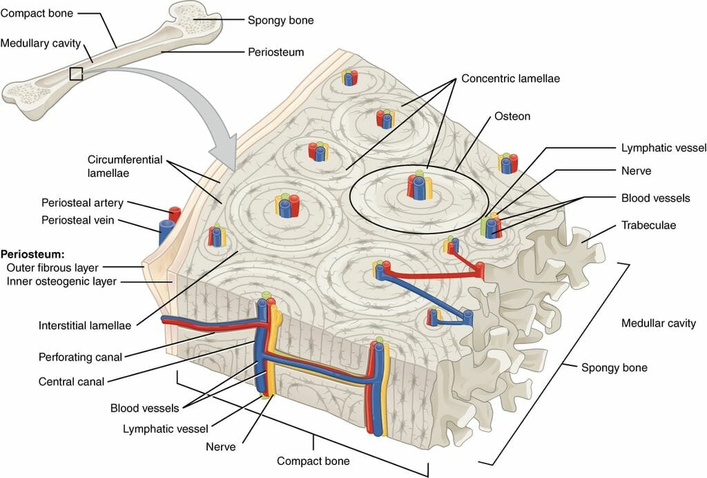
Compact Bone Structure Biology Dictionary
Compact Bone Cross Section

Compact Bone Diagram Labeled Compact Bone Labeling Production of
Transverse Section of Compact Bone ClipArt ETC

Compact bone location and function
What Is the Function of Compact Bone? (with pictures)
Skeletal Tissues Basicmedical Key

Anatomy microscopic structure of compact bone diagram (part 1

Histology of Bone step by step drawing of compact bone YouTube
What is compact bone tissue composed of? Socratic
Periosteum Osteons Perforating (Volkman’s) Canals Central Canals Lamellae (Layers Of Bony Material) Lacunae Lacunae [Type A Quote From The Document Or The Summary Of An Interesting Point.
These Oblique And Transverse Views Of The Diaphysis Of A Long Bone Show The Internal Structure Of Compact Bone.
Discover The Difference Between Spongy And Compact Bone, Learn About The Function Of Osteons, And Delve Into The Role Of Haversian Canals, Lacunae, And Volkmann Canals In Bone Health.
Spongy Or Cancellous Bone Histology.
Related Post: